Which to Choose CT Scan or X Ray, CT Scan Benefits


Which to Choose CT Scan or X Ray, CT Scan Benefits

The diagnostic centre is where the doctor performs all the tests to find the correct treatment for your disease. To find a nearby diagnostic centre, write on google diagnostic centre in Ludhiana.

To diagnose a person, many procedures are applied according to the symptoms of the person shown. 

CT scan should be done for complex problems like brain tumours etc. 

CT scan cost in Ludhiana is quite effective; no need to worry about pocket weight.

In this post, we will confer the CT scan procedure in detail. Whether you’re a doctor, patient, or just a student curious about CT scan procedure knowledge, this article will benefit all.

What is a CT scan:

A computed Tomography scan is often termed a CT scan. People often discuss their treatment and say the term CT scan in conversation. 

What is a CT scan? The scan imaging process helps doctors better understand the injury’s condition in your bones and tissues. It is a painless method that involves a series of 

X-rays to do the job. 

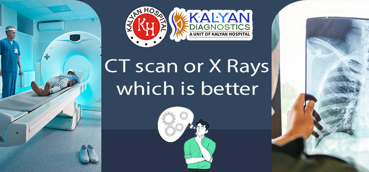
Difference between CT Scan and X Rays:

If a CT scan involves multiple X-rays at the job, why should we not use the X-ray machine? 

Why invest in another machine named CT Scan? 

A significant difference exists between the image produced by the X-ray machine and the CT Scan machine. The images formed by the X-rays cannot be seen clearly; the other body parts seem to be over, appearing in the image produced by the X-ray machine.

 But in the image produced by the CT Scan machine. Every organ can be seen clearly. This machine revolves around the body part to take multiple photos for an accurate and high-quality X-ray image of your body part or injury.

Be clear of the terms CT Scan and CAT Scan. Both are the same. CAT Scan stands for Computed Axial Tomography. 

Which parts can CT Scan show:

It can show you the best and clearer image of blood vessels, bones, muscles, and organs to be more precise for the treatment.

Problems detection of CT Scan : 

The CT scan can detect several problems. Some of the issues are listed below.:

Cancer: CTs are beneficial when discussing the most dangerous disease like cancer. CTs not only can confirm the cancer disease but also can check during the treatment whether the treatment is working or not.

Fractures: CT scan of a bone fracture can provide you with a more detailed structure of the bone injury than regular X-rays. And are incredibly accurate in the rupture of the wrist, ankle, legs, neck, etc.

Heart disease: A chest scan can give your doctor a detailed look at your heart condition. This can help in many cardiac-related diseases.

Brain injury: CT scan can provide the accurately detailed structure of your brain with any injury like bleeding or damaged nerves which cannot be seen in the x rays

Kidney stones:  are another common but hazardous problem among people nowadays. Many people don’t even know they have kidney stones that cannot be neglected. A CT scan helps to confirm if any person has a kidney stone. Kidney stones are detected easily in the CT scan.

Conclusion: 

There is no doubt that a CT scan is one of the most significant inventions of humanity. A CT scan helps detect many injuries that are not clear in X-rays.

It may be a symptom of any significant issue if you’re feeling back, neck, or kidney pain. Better it would help if you visited the Kalyan Diagnostic Center for proper treatment.

Is Mammography Testing the Right Choice for You?

Welcome to our blog on diagnostic centers and mammography tests. If you have ever wondered about these essential healthcare services, you are in the right place. In simple terms, diagnostic centers are places where doctors use special machines to check your body’s health. Mammography tests, specifically for women, help detect breast problems. We’ll explain everything in a way that’s easy to understand so you’ll be well-informed and prepared for your health journey. If you are finding the right Diagnostic center in Ludhiana, it is crucial for your well-being.

Breast Cancer Screening: Thermogram No Substitute for Mammogram

Breast cancer is a significant health concern, and early detection is crucial. It’s essential to recognize that although thermograms may appear as an option for mammograms, they are not a replacement. Let’s break down why:

  • Different Approaches

Thermograms use heat to detect changes in breast tissue. They capture temperature variations, which can be linked to potential issues. Mammograms, on the other hand, rely on X-rays to create detailed images of the breast. This method can reveal even the tiniest abnormalities.

  • Reliability Matters

Mammograms have long been the gold standard for breast cancer screening. They are trusted and have a well-established track record. Thermograms, while intriguing, are less reliable when it comes to detecting small tumors. Relying solely on thermograms could lead to missed opportunities for early intervention.

If you’re concerned about breast health, consider heading to a diagnostic center for a mammography test. It’s a decision that could ultimately save your life.

Mammography vs. Thermography

When considering breast health, two frequently mentioned diagnostic tools are mammography and thermography. But what are they, and how do they differ?

Mammography is like a special X-ray for breasts. It uses low-dose radiation to create detailed images. It’s great at detecting small lumps or tumors, especially in older women. But the radiation can be a concern for some.

Thermography, on the other hand, is all about heat. It captures infrared images of your breasts to detect temperature changes. It’s radiation-free and safe, making it suitable for younger women. But it might not spot small lumps as effectively as mammography.

Each has its pros and cons, and your doctor will help you decide which is best for you. Remember, early detection is key for breast health.

Advice for Patients Getting Breast Cancer Screening

Breast cancer screening is vital for your well-being, and it’s not as daunting as it may seem. The process is painless, quick, and non-invasive. When preparing for your mammography test, remember these three essential points:

  • Choose a reputable Center:

Select a diagnostic center with a solid reputation and experienced staff. Research and read reviews to ensure you’re in capable hands. 

  • Communication is Key:

Don’t hesitate to voice any concerns or questions to the technicians. They are there to assist and make you comfortable throughout the process.

  • Prepare and Stay Positive:

Dress in comfortable clothing, free from deodorant or lotion, for accurate results. Regardless of the outcome, stay positive and seek support from loved ones. Remember, regular screenings are your best defense against breast cancer, promoting a healthier and happier you.

Conclusion

When it comes to your well-being, our center stands out as the ideal choice for a Mammography Test in Ludhiana. By opting for our services, you ensure accuracy, professionalism, and a commitment to your health. Regular screening at Kalyan Diagnostics Centre is your best ally for a healthier future.

The importance of mammographic tests to examine breast issues

Mammographic tests are beneficial for examining the different issues of breasts. The mammographic tests used by the doctors for analyzing the breast tissue. Mammography is an X-ray imaging method used to examine the breast for the early detection of cancer and other breast diseases. It is used as both a diagnostic and screening tool.

What is a mammographic test?

Mammography is an X-ray procedure. The X-rays taken from a mammographic machine are known as mammograms. These mammograms are useful for a doctor to detect breast cancer at an early level. Regular mammograms can identify breast cancer. 

Reason for the mammograms 

A mammogram is a type of X-ray picture of the breast. Mammograms are helpful to examine the early signs of cancer of the breast. Daily basis mammograms are lucrative for finding breast cancer early. The human body takes a minimum of three years to notice the symptoms of breast cancer. The Mammography Test in Ludhiana is done at Kalyan Diagnostics.

Symptoms of breast cancer

People notice many symptoms if they are suffering from breast cancer. You may notice the following symptoms. 

  • You may feel lumps in the area of the breast and underarm. 
  • Feel swelling and thickening of the breast. 
  • Feel irritation and dimple breast skin. 
  • Redness and breaking skin of the nipple area and area of the breast.
  • Pain in any area of the breast.
  • Pain around the nipple area. 
  • Discharge from the nipple other than the milk, blood also includes.  
  • Notice the change in shape and size of the breast. 

If you notice any symptoms, then you should go to the best Diagnostic center in Ludhiana. Kalyan Diagnostics is considered the best diagnostic center. 

Cause of breast cancer 

Many things are responsible for breast cancer. Things such as age, hormonal medications, contraceptive pills and lifestyle factors can cause breast cancer. 

Natural factors

  • Age: Breast cancer occurs after the age of 50. The menstrual cycle stops after 50. Menopause is a reason for breast cancer after the age of 50.
  • Background history: if anyone in your family has ovarian cancer, it is at risk of breast cancer. There are the chances that you will also suffer from breast cancer because cells of cancer are also present in the genetic material. 
  • The dense tissue: The woman’s breast contains thousands of tiny glands. These are called lobules. Lobules are also known as milk glands. The tissue of glands is very dense. Women who have denser tissue are at the risk of breast cancer because it gives more space for the generation of cancer cells.

Hormones and hormonal medicines:  

  • Estrogen: An important hormone secretion named estrogen also causes breast cancer. The storage for eggs, called ovaries, starts producing estrogen at the start of puberty. It helps to regulate your periods. 
  • HRP: Hormonal replacement therapies can cause breast cancer if it is taken more than once a year. Except for vaginal estrogen, other HRP can be a reason for breast cancer. 
  • Contraceptive pills: Many women take contraceptive pills. The studies of the researcher show that the pills have a component that increases the risk of breast cancer. 

Lifestyle of a person 

  • Alcohol and drugs: Alcohol and drugs can increase the risk of breast cancer. 
  • Increase in weight: Obesity is a major problem. Many problems occur because of obesity. Obesity can be responsible for a high risk of breast cancer.
  • Radiation: People who are working in laboratories where X-ray and CT scans are done are at risk of breast cancer. These radiations are really dangerous for health. If you had an X-ray and CT done, then the chances of breast cancer are there. 

Diagnosis for breast cancer.

Doctors suggest you take different kinds of X-rays. With the help of these X-rays, doctors decide the treatment plan. 

  • Physical examination of breast
  • X-ray
  • Mammograms
  • Magnetic Resonance Imaging. 
  • CT scan 
  • Sample of cells of the breast
  • Ultrasound of breast

Treatment of breast cancer

There are two types of treatment according to the level of breast cancer: surgery and therapies. 

Surgery treatment

Surgeries are helpful in removing the lumps and cancer cells from the breast. The steps are as follows:

  • Lumpectomy: In this step, only cancer cells are removed
  • Mastectomy: Mastectomy is to remove the breast. It mostly performs in highly affected patients. 
  • Sentinel node biopsy: in this surgery, a limited number of lymph nodes are removed. 
  • Axillary lymph node dissection: Many lymph nodes are removed in this.
  • Removal of both breasts: Many women have breast cancer on one side only. They have to choose the removal of another breast because of genetic predisposition. 

Types of Therapies

Several types of therapies are beneficial for reducing the risk of breast cancer.

  • Radiation therapy
  • Chemotherapy
  • Hormonal therapy 
  • Immunotherapy

Breast cancer is a common problem in women in today’s era. The reason is an unhealthy diet and a busy lifestyle. To reduce the chances of breast cancer, females should eat the proper diet and live a healthy lifestyle.

जानें क्या है एमआरआई (MRI) जांच की सम्पूर्ण प्रक्रिया ?

एमआरआई का स्कैन करवाने की सलाह अक्सर डॉक्टर उन लोगों को देते है जिन्हे किसी तरह की अंधरुनि समस्या का सामना करना पड़ता है। तो अगर आपमें भी कोई ऐसी समस्या है जो आपकी परेशानी का कारण बनी हुई है तो इसके लिए आपको  एमआरआई (MRI) की जाँच जरूर करवानी चाहिए। इसके अलावा कब और किस समय इस जाँच का चयन करना चाहिए और क्या है इस जाँच की पूरी प्रक्रिया इसके बारे में भी आज के आर्टिकल में बात करेंगे, इसलिए एमआरआई (MRI) की सम्पूर्ण जानकारी के लिए आर्टिकल के साथ अंत तक बने रहें ;

क्या होता है एमआरआई (MRI) ?

एमआरआई (मैग्नेटिक रेसोनेंस इमेजिंग) इमेजिंग जांच का एक प्रकार है। इसमें शरीर के अंदर की उच्च गुणवत्ता वाली, विस्तृत तस्वीरें बनाने के लिए एक बड़े चुंबक, रेडियो तरंगों और कंप्यूटर का उपयोग किया जाता है। एमआरआई जांच में दर्द नहीं होता। इसलिए अगर आप इस जाँच का चयन अपने या अपने बच्चे के लिए करते है तो दोनों को ही दर्द का आभास नहीं होगा।

क्या है एमआरआई (MRI) जांच ?

कई बार गंभीर और आंतरिक शारीरिक समस्या होने पर डॉक्टर अक्सर रोगी को एमआरआई जांच करवाने के लिए कहते है। जिसमें शरीर के अंदर क्या हो रहा है इस बारे में जांच की जाती है और इसके बाद रोगी को उचित उपचार प्रदान किया जाता है। चिकित्सा के क्षेत्र में एमआरआई का बहुत महत्व है जो कि रोगी को उचित उपचार प्रदान करवाने में काफी सहायक है, इसलिए इस के बारे में विशेष जानकारी होना आवश्यक है।

एमआरआई से क्या पता लगता है ?

  • आपके मस्तिष्क और आसपास के तंत्रिका ऊतक का पता लगना।
  • आपकी छाती और पेट के अंग, जिसमें आपका हृदय, लीवर, पित्त पथ, गुर्दे, प्लीहा, आंत्र, अग्न्याशय और अधिवृक्क ग्रंथियां शामिल है।
  • स्तन ऊतक के बारे में एमआरआई स्कैन से पता लगना।
  • आपकी रीढ़ की हड्डी का पता लगना।
  • आपके मूत्राशय और प्रजनन अंगों सहित श्रोणि अंग का पता लगना। 
  • रक्त वाहिकाओ के बारे में जानकारी आदि।

अंतर क्या है एमआरआई (MRI) और सीटी (CT) स्कैन में ?

  • मैग्नेटिक रेज़ोनेंस इमेजिंग (MRI) आपके शरीर के अंदर की छवियों को बनाने के लिए मैग्नेट, रेडियो तरंगों और एक कंप्यूटर का उपयोग करता है। जहां एमआरआई अंधरुनि ऊतकों के बारे में जानकारी देता है, वही लुधियाना में MRI का खर्च 1,500 से 2500 तक का आ सकता है।
  • जबकि कंप्यूटेड टोमोग्राफी (सीटी) एक्स-रे और कंप्यूटर का उपयोग करता है। और लुधियाना में CT स्कैन का खर्च निर्भर करता है की आप शरीर के किस तरह के अंगों का स्कैन करवाते है।

क्या एमआरआई जांच को सुरक्षित माना जा सकता है ?

  • एक MRI scan आम तौर पर सुरक्षित माना जा सकता है और उचित सुरक्षा दिशानिर्देशों का पालन करने पर औसत व्यक्ति के लिए कोई जोखिम नहीं होता। 
  • एमआरआई मशीनों से निकलने वाला मजबूत चुंबकीय क्षेत्र आपके लिए हानिकारक नहीं है, लेकिन इससे प्रत्यारोपित चिकित्सा उपकरण खराब हो सकते है।

सीटी व एमआरआई जांच के लिए बेस्ट हॉस्पिटल व सेंटर !

  • अगर आपने उचित दाम पर सीटी व एमआरआई स्कैन को करवाना चाहते है तो इसके लिए आपको कल्याण डायग्नोस्टिक सेंटर का चयन करना चाहिए।

निष्कर्ष :

जैसा की आपको पता ही चल गया होगा की किस तरह से हम एमआरआई स्कैन की मदद से शरीर के अंधरुनि परेशानियों के बारे में जानकारी हासिल कर सकते है और इसके बाद अपनी बीमारियों का इलाज आसानी से किसी अनुभवी डॉक्टर से करवा सकते है।

भारत में क्या है एमआरआई (MRI) स्कैन की लागत व पूरी प्रक्रिया ?

कई बार व्यक्ति को कुछ ऐसी बीमारियां हो जाती है कि उसे पता ही नहीं चलता है की वो किस तरह की बीमारी का सामना कर रहा है पर ऐसे में एमआरआई (MRI) स्कैन एक बेहतरीन विकल्प माना जाता है अंदरूनी बीमारी को जानने में तो अगर आप भी किसी तरह की बीमारी का सामना कर रहे और वो बीमारी आपको काफी परेशान कर रही है तो उससे निजात पाने के लिए आपको एमआरआई स्कैन का चयन करना चाहिए ;

क्या है एमआरआई (MRI) स्कैन ?

  • चुंबकीय अनुनाद इमेजिंग यानि “मैग्नेटिक रेज़ोनेंस इमेजिंग” एक चिकित्सा इमेजिंग तकनीक है जो कि आपके शरीर में अंगों और ऊतकों की विस्तृत छवियां बनाने के लिए चुंबकीय क्षेत्र और कंप्यूटर से उत्पन्न रेडियो तरंगों का उपयोग करती है। वहीं यह एक्स-रे (विकिरण) का उपयोग नहीं करता है।
  • दूसरी और अधिकांश एमआरआई मशीनें बड़ी, ट्यूब के आकार की चुम्बक होती हैं। वही जब आप MRI मशीन के अंदर लेटते हैं, तो चुंबकीय क्षेत्र अस्थायी रूप से आपके शरीर में पानी के अणुओं को पुन: बनाता है।

 एमआरआई (MRI) स्कैन करवाने से क्या पता चलता है ?

  • मस्तिष्क, रीढ़, जोड़ों, हृदय, लीवर, और कई अन्य अंग जैसे शरीर के कोमल-ऊतक संरचनाओं की एमआरआई (MRI) चित्रों के द्वारा ज्यादा सटीक रूप से पहचाना जा सकता है। कैंसर, हृदय की बीमारी, मांसपेशियों और हड्डियों की बीमारियों, और कई अन्य बीमारियों का डायग्नोसिस एमआरआई स्कैन से किया जाता है।

 एमआरआई (MRI) स्कैन का उपयोग कहां किया जाता है ?

  • मस्तिष्क और रीढ़ की हड्डी की असामान्यताएं को जानने के लिए।
  • शरीर के विभिन्न भागों में द्रव्यमान, ट्यूमर, अल्सर और अन्य विसंगतियों की उपस्थिति को जानना।
  • पेट में हृदय, यकृत और अन्य अंगों से संबंधित स्थितियों को जानना।
  • फाइब्रॉएड और एंडोमेट्रियोसिस जैसी महिलाओं में पेल्विक दर्द से संबंधित स्थितियों को जानना।
  • बांझपन के मामले में महिलाओं में संदिग्ध मूत्र असामान्यताओं का मूल्यांकन करना।

आप एमआरआई से भी इन परेशानियों की जाँच करवा सकती है या आप लुधियाना में CT स्कैन का खर्च कितना आएगा, इसके बारे में जान के भी इसकी जाँच सीटी स्कैन से करवा सकते है।

एमआरआई (MRI) स्कैन को करवाने में खर्चा कितना आता है ?

  • सिटी स्कैन, एमआरआई की जांच के लिए मरीज से 500 से लेकर 8 हजार या उससे ज्यादा की फीस ली जाती है। हालांकि ये टेस्ट अभी कॉन्ट्रैक्ट बेस पर मशीनें लगाकर करवाई जा रही है।
  • इसके अलावा मरीज़ के अंगों पर भी इसका खर्चा निर्भर करता है की वो शरीर के किस अंग की जाँच करवाने जा रहा है। वही लुधियाना में MRI का खर्च जगह और हॉस्पिटल कहाँ स्थित है इस पर भी पड़ता है।

एमआरआई (MRI) स्कैन करवाने के लिए बेस्ट हॉस्पिटल व सेंटर ?

  • अगर आपको अपने शरीर के अंधरुनि अंगों की जाँच अच्छे से करवानी है तो इसके लिए आप हमारे  डायग्नोस्टिक्स सेंटर का चयन कर सकती है।

क्या है एमआरआई (MRI) स्कैन की प्रक्रिया ?

  • रोगी को स्कैन के दौरान अस्पताल का गाउन पहनने की आवश्यकता होती है। साथ ही, स्कैन के दौरान आभूषण या धातु पहनने की अनुमति नहीं है।
  • एमआरआई स्कैन एक गैर-इनवेसिव और दर्द रहित प्रक्रिया है। और अवधि स्कैन किए जा रहे शरीर के हिस्से के आकार और कितनी छवियों को लेने की आवश्यकता के आधार पर 15 मिनट से एक घंटे या उससे अधिक तक समय लग सकता है।

निष्कर्ष :

उम्मीद करते है की आपको पता चल गया होगा की आपको कब और किस समय एमआरआई (MRI) स्कैन करवाना चाहिए।

Decoding the Differences between CT Scan vs MRI

Imaging technologies are critical in delivering insights into the human body in the area of medical diagnosis. CT scans (Computed Tomography) and MRI (Magnetic Resonance Imaging) are two regularly utilized imaging procedures. While both CT scans and MRI have revolutionized diagnostic medicine, they differ in key ways, including the technology used, the sorts of pictures produced, and their applications. Understanding the distinctions between these two imaging modalities is critical for both patients and healthcare practitioners. In this blog, we will decipher the differences between CT scans and MRIs, providing light on their distinct qualities, benefits, and applications. Whether you are looking for a diagnostic center or simply want to understand more about CT scan cost in Ludhiana, this blog is for you.

Understanding CT Scan:

A CT scan, otherwise called figured tomography, uses X-beams and a PC to make point-by-point cross-sectional pictures of the body. It gives an exhaustive perspective on bones, organs, and tissues, permitting healthcare experts to detect irregularities, recognize wounds, and analyze the scope of ailments. CT scans are especially valuable for assessing head wounds, recognizing growths, evaluating interior dying, and looking at the chest, mid-region, and pelvis.

Investigating MRI:

MRI, or attractive reverberation imaging, utilizes a powerful attractive field and radio waves to produce exceptionally itemized pictures of the body’s structures. Not at all like CT scans, MRI doesn’t include radiation, making it a more secure choice for specific people. MRI is known for its capacity to give multifaceted pictures of delicate tissues, like the mind, spinal string, joints, and organs. It is normally used to analyze conditions like cerebrum growths, spinal line wounds, joint problems, and cardiovascular infections.

Distinguishing Factors:

  1. Innovation and Picture Arrangement:

CT scans depend on X-beam innovation to deliver pictures, while MRI utilizes attractive fields and radio waves. This key distinction in innovation impacts the kind of data each imaging procedure catches. CT scans succeed at featuring bone structures and detecting thick tissues, while MRI offers the uncommon perception of delicate tissues and definite physical structures.

  1. Radiation Openness:

One huge factor to consider is radiation openness. CT scans include openness to ionizing radiation, which conveys likely dangers, especially with rehashed openness. Then again, MRI doesn’t utilize radiation, making it a favored choice for people who might be more delicate to radiation or require different imaging meetings.

  1. Picture Lucidity and Difference:

CT scans are known for their excellent lucidity and capacity to envision bone structures and thick tissues with high differences. They are especially effective in detecting fractures, growths, and calcifications. MRI, then again, gives definite pictures of delicate tissues, offering better differentiation for recognizing healthy and strange tissues.

Conclusion:

Understanding the differences between CT scans and MRIs is pivotal in deciding the most suitable diagnostic imaging method for your particular clinical requirements. By talking with a trusted diagnostic centre in Ludhiana, like Kalyan Diagnostics you can settle on informed conclusions about which imaging methodology, CT scan or MRI, is the most ideal for your specific circumstance. Have confidence, both CT scans and MRI are powerful devices in the possession of gifted clinical experts, empowering exact judgments and effective therapy making arrangements for a great many ailments.

जानिए 2D इको टेस्ट हार्ट ब्लॉक का पता लगाने में कैसे मददगार है ?

2D इकोकार्डियोग्राफी टेस्ट हार्ट की समस्याओं का पता लगाने में काफी मददगार साबित होते है। लेकिन इस टेस्ट को लेकर बहुत से लोगों के मन में ये सवाल होता है की क्या उन्हे इस टेस्ट का चयन अपने अच्छे स्वास्थ्य की समस्याओं को जानने के लिए करना चाहिए या नहीं, तो इसको लेकर आज एक लेख में चर्चा की जाएगी ;

2D इको टेस्ट क्या है ?

  • 2डी इकोकार्डियोग्राफी और कलर डॉप्लर एक ऐसा टेस्ट है जो दिल की सोनोग्राफी जितना ही अच्छा है। इन परीक्षणों में, आप हृदय को अंदर से देख सकते है जैसे हृदय की मांसपेशियां, वाल्व, रक्त प्रवाह, कक्ष का फैलाव, और हृदय कार्य करता है लेकिन आपको हृदय में ब्लॉक नहीं दिखाई देते है।
  • वहीं इकोकार्डियोग्राफी परीक्षण में आपके दिल की जीवंत छवियों को बनाने के लिए ध्वनि तरंगों का उपयोग किया जाता है। छवि बना कर दिल की स्थिति जानना इकोकार्डियोग्राम है। यह परीक्षण आपके डॉक्टर को यह देखने की अनुमति देता है कि आपका दिल और उसके सारे तत्व कैसे काम कर रहे है। दिल की कोई भी कमी जो विशेष कर दिल की अंदुरुनी होती है उन्हें इको से ढूंढा जा सकता है।

क्यों किया जाता है 2D इको टेस्ट ?

  • डॉक्टर आपके दिल की संरचना को देखने के लिए एक इको टेस्ट करवाने की सलाह आपको देते है और मूल्यांकन करते है कि आपका दिल कितनी अच्छी तरह काम कर रहा है। 
  • वहीं इको टेस्ट निम्नलिखित स्थितियों के निदान के लिए किया जाता है :-
  • हृदय में प्रवेश करने या छोड़ने वाली बड़ी रक्त वाहिकाओं से संबंधित समस्या का सामना करने वाले इसका चयन करते है। 
  • पेरीकार्डियम से संबंधित मुद्दे, यानी दिल की बाहरी के बारे में जानने के लिए इसका चयन किया जाता है। 
  • ह्रदय कक्षों के बीच किसी भी छेद की मौजूदगी का होना। 
  • ह्रदय के कक्षों में रक्त के थक्कों के जमने की मौजूदगी का होना। 
  • समय के साथ हृदय रोग की निगरानी को करना।
  • 2D इको टेस्ट करवाने के लिए आपको लुधियाना में बेस्ट रेडियोलॉजिस्ट का चयन करना चाहिए।

2D इको टेस्ट कैसे किया जाता है ?

  • यह एक साधारण परीक्षण है जो एक क्लिनिक में आउट पेशेंट के आधार पर किया जा सकता है, जहां आप मेज पर लेटते है, इसमें मुश्किल से 2 मिनट लगते है और डॉक्टर हृदय की कार्यप्रणाली को बता सकते है, लेकिन पुराने दिल के दौरे की पुष्टि करने के लिए यह एक अच्छा परीक्षण होगा दिल का दौरा यह भविष्य के दिल के दौरे की भविष्यवाणी नहीं कर सकता।
  • शुरू करने के लिए, रोगी को लेटने और कमर से ऊपर तक कपड़े निकालने का निर्देश दिया जाता है।
  • फिर उसे एक पेपर शीट या एक बेड शीट का उपयोग करके कवर किया जाएगा, जो पूर्वकाल छाती को उजागर करेगा।
  • इलेक्ट्रोड को रोगी के शरीर पर रखा जाएगा।
  • उसे गहरी साँस लेने या अपनी तरफ से झूठ बोलने से अपनी स्थिति बदलने के लिए कहा जा सकता है।
  • प्रक्रिया शुरू होने से पहले छाती पर एक विशेष इको-जेल लागू किया जाएगा।

2D इको टेस्ट का उद्देश्य क्या है ?

  • दिल के वाल्व का खराब होना। 
  • जन्मजात हृदय दोष की समस्या।  
  • दिल के दौरे के बाद दिल को संभावित नुकसान होना। 
  • दिल की सूजन का कोई भी लक्षण (पेरिकार्डिटिस)।
  • दिल की थैली के आसपास कोई तरल पदार्थ का जमा होना। 
  • हृदय में नर्म ध्वनि को ठीक करना।

2D इको टेस्ट की तैयारी कैसे करें ?

यह एक बहुत ही सुरक्षित परीक्षा है, जिसे विशेष रूप से व्यवस्थित वातावरण में प्रशिक्षित सोनोग्राफर के तहत किया जाना चाहिए। 

और इसमें किसी भी विशेष तैयारी की आवश्यकता नहीं है, लेकिन मधुमेह के रोगियों को सलाह दी जाती है कि वे इस प्रक्रिया से पहले डॉक्टर से परामर्श करें क्योंकि यदि कार्डियोलॉजिस्ट ऐसा सुझाते है, तो दवा की खुराक में संभावित परिवर्तन हो सकता है।

2D इको टेस्ट से हमें किन महत्वपूर्ण बातों के बारे में पता चलता है ?

  • इकोकार्डियोग्राम हृदय की दीवारों के मोटे होने या हृदय के कक्षों के बढ़ने के कारण हृदय के आकार में किसी भी परिवर्तन को प्रभावी ढंग से निर्धारित कर सकता है। हृदय के आकार में परिवर्तन का कारण बनने वाले मुख्य कारणों में उच्च रक्तचाप का क्षतिग्रस्त या कमजोर हृदय वाल्व का होना शामिल है।
  • इको रिपोर्ट इजेक्शन फ्रैक्शन और कार्डियक आउटपुट का प्रतिशत भी देती है (यानी एक मिनट में जितना रक्त पंप किया जाता है)। अपर्याप्त पंपिंग शक्ति वाला हृदय, हृदय की विफलता का कारण बन सकता है।
  • एक इकोकार्डियोग्राम की मदद से, डॉक्टर हृदय की मांसपेशियों को किसी भी नुकसान की पहचान कर सकता है, जो हृदय की सामान्य पंपिंग को प्रभावित कर सकता है।
  • एक इकोकार्डियोग्राम यह भी बता सकता है कि रक्त के रिसाव को रोकने के लिए आपके हृदय के वाल्व खुले और पूरी तरह से बंद है या नहीं।
  • एक इकोकार्डियोग्राम प्रमुख रक्त वाहिकाओं और हृदय के बीच असामान्य कनेक्शन, हृदय कक्षों से संबंधित मुद्दों और जन्म के समय मौजूद हृदय में किसी भी दोष की पहचान कर सकता है।

2D हार्ट ब्लॉक इको टेस्ट का खर्चा कितना आता है ?

  • लुधियाना में 2D इको टेस्ट का खर्च 1000 से 3000 रुपये के बीच हो सकता है। वहीं इसका खर्च निर्भर करता है की टेस्ट करने वाले रेडियोलॉजिस्ट का अनुभव कितना पुराना है और साथ ही हॉस्पिटल किस जगह पर स्थित है। 
  • इसके अलावा आप चाहें तो इस टेस्ट को कल्याण डायग्नोस्टिक्स हॉस्पिटल से भी करवा सकते है क्युकी यहां पर मरीज का टेस्ट अनुभवी डॉक्टरों के द्वारा किया जाता है और टेस्ट को करने के लिए आधुनिक या नवीनतम उपकरणों का भी प्रयोग किया जाता है।  

ध्यान रखे :

हार्ट अगर सुचारु रूप से चलेगा तभी जाकर हम किसी भी तरह की समस्या से खुद का बचाव आसानी से कर सकते है, और हार्ट को ठीक तरीके से हम किस तरह से चला सकते है इसके बारे में हम उपरोक्त बता चुके है।

निष्कर्ष :

हार्ट संबंधी समस्याओं के बारे में जानने के लिए किस तरह से 2D इको टेस्ट मददगार है, इसके बारे में हम आपको उपरोक्त्त बता ही चुके है। तो अगर आपको अपने हार्ट में किसी भी तरह की समस्या नज़र आए तो इससे बचाव के लिए आपको इस टेस्ट का चयन करना चाहिए।

क्‍या है डॉप्‍लर टेस्‍ट (Doppler Test) और प्रेगनेंसी में इस टेस्ट की जरूरत क्‍यों होती है ?

गर्भावस्था एक बहुत ही नाजुक स्टेज है महिलाओं की जिंदगी में। इस स्टेज में महिलाओं को अपने साथ-साथ अंदर पल रहें बच्चे का भी अच्छे से ध्यान रखना होता है और इस ध्यान को वह समय-समय पर होने वालें टेस्टों को करवा कर रख सकती है और उन्ही टेस्टों में एक टेस्ट डॉप्‍लर टेस्‍ट या स्‍कैन या अल्‍ट्रासाउंड है। हालांकि, कई गर्भवती महिलाओं काे इस बात की जानकारी नहीं होती है कि डॉप्‍लर टेस्‍ट क्‍यों करवाया जाता है और इससे क्‍या पता चलता है। तो वहीं अगर आप इस टेस्ट को करवाना चाहते है तो इसके बारे में जानकारी हासिल करने के लिए लेख के साथ अंत तक बने रहें ;

क्या है डॉप्‍लर टेस्‍ट (Doppler Test) ?

  • डॉप्‍लर स्‍कैन एक तरह का रेगुलर अल्‍ट्रासाउंड स्‍कैन ही होता है और इसमें हाई फ्रीक्‍वेंसी साउंड वेव्‍स का इस्‍तेमाल किया जाता है। नॉर्मल अल्‍ट्रासाउंड की तरह ही इसकी प्रक्रिया होती है और इसमें आप कंप्‍यूटर पर गर्भस्‍थ शिशु की तस्‍वीर देख सकते है।
  • रेगुलर अल्‍ट्रासाउंड से डॉप्‍लर स्‍कैन इसलिए अगल होता है, क्‍योंकि ये रक्‍त वाहिकाओं में रक्‍त के प्रवाह, खून के प्रवाह की गति, दिशा और खून के थक्‍के के बारे में भी बताता है। आजकल अधिकतर अल्‍ट्रासाउंड में इनबिल्‍ट डॉप्‍लर फीचर होता है और दोनों ही स्‍कैन एक साथ किए जा सकते है।

डॉप्‍लर टेस्‍ट (Doppler Test) क्यों किया जाता है ?

  • यदि पेट में जुड़वां बच्‍चे हों, शिशु रीसस एंटीबॉडीज से प्रभावित हो, शिशु का स्‍वस्‍थ विकास न हो रहा हो, पहली प्रेगनेंसी में शिशु का आकार छोटा रहा हो, पहले डिलीवरी से मिसकैरेज हुआ हो या डिलीवरी के वक्‍त बच्‍चा मर गया हो, डायबिटीज या हाई ब्‍लड प्रेशर से ग्रस्‍त प्रेगनेंट महिला को, जिनका बीएमआई (बॉडी मास इंडेक्‍स) कम या ज्‍यादा हो तो इन स्थितियों में डॉप्‍लर टेस्‍ट किया जाता है।
  • प्‍लेसेंटा ठीक तरह से काम कर रहा है या नहीं, और इसे शिशु को पर्याप्‍त ऑक्‍सीजन और पोषण मिल पा रहा है या नहीं, इस बात का पता लगाने के लिए भी डॉप्‍लर स्‍कैन किया जाता है।
  • डॉप्‍लर स्‍कैन से डॉक्‍टर को पता चलता है कि क्‍या शिशु की सेहत को ठीक करने के लिए कोई कदम उठाने या जल्‍दी डिलीवरी करवाने की जरूरत तो नहीं है।
  • वहीं डॉप्‍लर स्‍कैन को करवाने के लिए आपको लुधियाना में बेस्ट रेडियोलॉजिस्ट का चयन करना चाहिए।

क्या प्रेगनेंसी में महिलाएं डॉप्‍लर टेस्‍ट (Doppler Test) करवा सकती है ?

  • जी हां, गर्भवती महिला के लिए डॉप्‍लर स्‍कैन सुरक्षित है, लेकिन आपको एक प्रशिक्षित अल्‍ट्रासाउंड डॉक्‍टर से ही ये स्‍कैन करवाना है। इस अल्‍ट्रासाउंट से शिशु की सेहत के बारे में पूरी जानकारी पाने में मदद मिलती है।
  • आमतौर पर प्रेगनेंसी की तीसरी तिमाही यानी 36 और 40वें सप्‍ताह में ग्रोथ स्‍कैन के साथ डॉप्‍लर स्‍कैन किया जाता है। हालांकि, अगर गर्भावस्‍था में कोई परेशानी हो तो इससे पहले भी डॉप्‍लर टेस्‍ट करवाया जा सकता है।
  • तीसरी तिमाही से पहले डॉप्‍लर टेस्‍ट शिशु में जेनेटिक और कार्डिएक विकारों का पता लगाने के लिए किया जाना चाहिए। इस समय अल्‍ट्रासाउंड डॉक्‍टर को 5 से 10 मिनट से ज्‍यादा समय इस टेस्‍ट में नहीं लगाना चाहिए।
  • डॉप्‍लर और कलर स्‍कैन में थर्मल इंडेक्‍स थोड़ा अधिक होता है लेकिन फिर भी इसकी कम डोज सुरक्षित मानी जाती है। कम से कम समय के लिए आप इस स्‍कैन को कर सकते है। अधिकतर मामलों में डॉप्‍लर स्‍कैन सिर्फ कुछ मिनट के लिए ही किया जाना चाहिए।

कैसे किया जाता है डॉप्‍लर टेस्‍ट (Doppler Test) ?

  • नॉर्मल अल्‍ट्रासाउंड की तरह ही डॉप्‍लर स्‍कैन किया जाता है। और इसमें प्रेगनेंट महिला को टेबल पर लिटाकर पेट पर वॉटर बेस जैल लगाया जाता है। अब ट्रांसड्यूसर को पेट पर लगाया जाता है और इससे आप कंप्‍यूटर की स्‍क्रीन पर उसी समय अपने शिशु की तस्‍वीरें देख सकती है। 
  • इस स्‍कैन में केवल कुछ मिनट का समय लगता है और इसमें दर्द भी नहीं होता है।
  • ब्‍लड फ्लो को चेक करने के ल‍िए स्‍कैनर ध्‍वन‍ि तरंगों का इस्‍तेमाल भी किया जाता है। जबकि बात करें सामान्‍य अल्‍ट्रासाउंड की तो इसमें ब्‍लड फ्लो का पता नहीं लगाया जा सकता है। 

डॉप्‍लर टेस्‍ट (Doppler Test) से शिशु के बारे में क्या जानकारी मिलती है ?

  • श‍िशु की हलचल के बारे में पता चलता है।
  • प्रेगनेंसी में ब्‍लड फ्लो कैसा है, इसकी जानकारी म‍िलती है।
  • गर्भस्‍थ श‍िशु की बीमारी के बारे में भी पता चलता है। 
  • जुड़वां बच्‍चों की स्‍थि‍त‍ि के बारे में भी जानकारी मिलती है।

डॉप्‍लर टेस्‍ट करवाने से पहले लुधियाना में कलर डॉपलर टेस्ट का खर्च कितना आता है, इसके बारे में जरूर जानकारी हासिल करें।

क्या डॉप्‍लर टेस्‍ट (Doppler Test) का कोई जोखिम भी है ?

  • डॉप्‍लर टेस्‍ट वाला अल्ट्रासाउंड सुरक्षित है और इसे गर्भवती रोगियों की पसंद का इमेजिंग तरीका माना जाता है। 
  • मानक बी-मोड और एम-मोड अल्ट्रासाउंड तरंगों की तुलना में, डॉपलर तरंगों को उच्च ध्वनिक आउटपुट की आवश्यकता होती है और भ्रूण के ऊतकों को थर्मल क्षति का एक उच्च सैद्धांतिक जोखिम प्रदान करता है। 
  • जब भी संभव हो भ्रूण के मूल्यांकन के लिए डॉपलर तरंगों की तुलना में बी-मोड और एम-मोड अल्ट्रासाउंड तरंगों की सिफारिश की जाती है।

डॉप्‍लर टेस्‍ट (Doppler Test) के लिए बेस्ट हॉस्पिटल !

  • अगर आप डॉप्‍लर टेस्‍ट करवाने के बारे में सोच रहीं है, तो इसके लिए आपको कल्याण डायग्नोस्टिक्स हॉस्पिटल का चयन करना चाहिए और इस हॉस्पिटल में अनुभवी रेडियोलॉजिस्ट के द्वारा इस टेस्ट को किया जाता है। 
  • वहीं इस टेस्ट को करने के दौरान जिस सावधानी को बरतना चाहिए वो इस हॉस्पिटल में बखूबी निभाया जाता है। इसके अलावा यहाँ पर किफायती दाम में इस टेस्ट को किया जाता है और इस टेस्ट को करने के लिए आधुनिक उपकरणों का प्रयोग किया जाता है।

निष्कर्ष :

डॉप्‍लर टेस्‍ट का चयन गर्भवती महिलाओं को क्यों करना चाहिए उम्मीद करते है, की आपको पता चल गया गया होगा। वहीं इस टेस्ट के दौरान किन बातो को ध्यान में रखना चाहिए और इस टेस्ट को किन डॉक्टरों के द्वारा करवाया जाना चाहिए, इसके बारे में भी हम आपको उपरोक्त बता चुके है। बस ध्यान रखें इसको करवाने के लिए किसी अनुभवी रेडियोलॉजिस्ट का ही चयन आपको करना चाहिए।