Guide to Lumbar MRI Scan: Purpose, Procedure, and Risks


Guide to Lumbar MRI Scan: Purpose, Procedure, and Risks

Most patients with low back discomfort with nerve root compression benefit from MRI Lumbar spine imaging. When it comes to identifying nerve damage brought on by spondylolisthesis, spinal stenosis, or intervertebral disc herniation, the scan is quite sensitive. Magnetic resonance imaging is the most effective imaging technique for examining and diagnosing such lumbar area issues. 


What is the definition of the MRI Lumbar spine imaging?

Patients experiencing low back pain that is related to nerve root compression are the primary candidates for MRI lumbar spine. Spondylolisthesis, spinal stenosis and intervertebral disc herniation can all cause nerve injury that the very sensitive scan can detect. Lumbar magnetic resonance imaging is the most effective technique for assessing and identifying such issues. Book an appointment with the best diagnostic center in Ludhiana to get a quality MRI. 


Why is MRI lumbar spine imaging done? 

The lumbar MRI spine imaging can check all the bone problems related to the spinal cord. 

  • Back pain accompanied by fever. 
  • Congenital disabilities affect your spine.
  • Injury to your lower spine.
  • Persistent or severe lower back pain.
  • Multiple sclerosis.
  • Problems with your bladder.
  • Signs of brain and spinal cancer. 
  • Weakness, numbness and other issues with your legs.


Preparation for the MRI lumbar spine imaging

Before the scan, your doctor will ask you to remove all jewelry and body piercings and change into a hospital gown. MRI magnets can occasionally attract metals. The preparation for scan is as following: 

  • Artificial heart valves
  • Clips
  • Implants
  • Pins
  • Plates
  • Prosthetic joints or limbs
  • Screws
  • Staples
  • Stents


Benefits of the MRI lumbar spine imaging

There are several benefits of the MRI lumbar spine imaging. 

  • Detailed Visualization: Magnetic Resonance Imaging offers pictures of the lumbar spine, encompassing the muscles, nerves, spinal cord, discs, vertebrae, and other soft tissues.
  • Non-Invasive: Magnetic resonance imaging is a non-invasive method that does not employ ionizing radiation, unlike CT or X-ray scans.
  • Multi-Plane Imaging: Magnetic Resonance Imaging enables imaging in several planes, offering various perspectives of the lumbar spine.
  • Soft Tissue Differentiation: Magnetic Resonance Imaging helps distinguish between various soft tissue types. This is essential for diagnosing infections, tumors, spinal stenosis, herniated discs, and spinal cord and nerve root health.
  • Functional Information: Diffusion-weighted imaging and functional MRI are two modalities that can shed light on the spinal cord and nerves’ current state of function.
  • Identification of Pain Source: An MRI scan can assist in locating potential pain sources, such as herniated discs, degenerative changes, or anomalies in the spine, for individuals who have chronic lower back pain.
  • Pre-Surgical Planning: MRI scans can give doctors vital information regarding the type and location of anomalies if surgery is considered a therapeutic option.


Risk factor of MRI lumbar spine imaging

Unlike an X-ray or CT scan, an MRI does not require ionizing radiation. It’s seen to be a safer substitute, particularly for expectant mothers and developing kids. Although they do occasionally occur, adverse effects are incredibly uncommon. The radio waves and magnets utilized in the scan have not yet been associated with known adverse effects.

Individuals with metal implants run specific hazards. The magnets used in an MRI can cause implanted screws or pins to move around in your body, or they can cause issues with pacemakers.

The diagnostic center does the MRI with a prescription given by a well-qualified and experienced doctor. Book an appointment with Kalyan Diagnostics for better-quality scans. It is also one of the best centers for MRI scans. MRI Cost in Ludhiana is only 3500-7000 bucks.

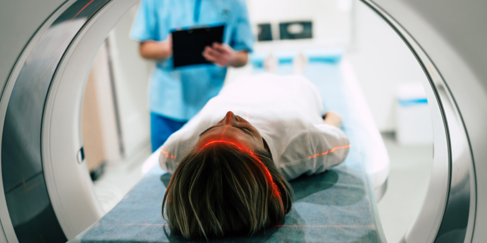
MRI Scans For Breast: What You Need To Know?

An MRI or Magnetic Resonance Imaging is a medical imaging test technique. It is highly used for detecting diseases as it is non-invasive and produces detailed images of the body’s internal structure. It provides details about organs, muscles, bones and blood vessels. It uses radio waves and magnetic waves, which help to create detailed images of the body. An MRI scan is also used to detect abnormalities in the breast. A reputed Diagnostic centre in Ludhiana provides MRI scans for breast at affordable cost. 

MRI Scan For Breast

When your doctor suggests an MRI scan for the breast, it is usually done to diagnose and examine the breast tissues for abnormalities such as cysts, tumours, and similar changes. Doctors typically suggest MRI in conjunction with other imaging testing techniques such as mammography and ultrasounds to get a detailed evaluation of the breast. Doctors can recommend MRI scans, although it may not be required for every woman having breast malignancy. Some tumours can be missed during a mammogram; hence, MRI scans are recommended for clear and detailed examination, and MRI cost in Ludhiana is reasonable. 

MRI Process

If you go for an MRI scan of your breast, the process will involve the following steps:

  • Preparation – As MRI machines create a strong magnetic field to get clear images of the body, you will be asked to remove any metal object and change it into a gown. If you have any metal implants or devices in your body, you must inform the staff because they can interfere with the MRI. 
  • Contrast Agent – A gadolinium compound dye will be injected into your veins to increase the visibility of certain tissues and blood vessels.  
  • Positioning – You are required to lie face down. To scan the breasts without compressing them, they are hung into an aperture. A good Diagnostic centre in Ludhiana will provide a padded table to ensure comfort. Also, to prevent you from shifting, extra cushions can be employed. 
  • Scan – As mentioned above, MRI uses strong radio and magnetic waves for detailed scanning and imaging. You will be required to stay still and quiet during the scan.
  • Duration – An MRI scan typically takes 30 to 60 minutes, depending on the type of scan or data required. 
  • Interpretation – Radiologists interpret MRI scans; they study the scan results and provide a detailed report to your healthcare provider. 

When Or Why Is an MRI Scan Required?

  • It is required for women with dense breast tissues as, in such cases, mammography can be less effective.
  • For detecting early breast cancer, which other tests may not have detected, you do not need to worry, as MRI cost in Ludhiana is quite budget-friendly.
  • For detailed examination of breast abnormalities and implants detected during mammography or ultrasound. 
  • They are used for screening patients or individuals who are at high risk due to a family history of cancer or genetic mutations predisposing to breast cancer. 
  • For women who took radiation therapy for the treatment of diseases such as Hodgkin’s disease between the ages of 10 and 30.

Conclusion

MRI scans are used to diagnose and detect problems that may have been invisible or overlooked by other tests such as mammography and ultrasound. However, you do not need to be scared as it is for a clear and detailed examination. An excellent diagnostic centre in Ludhiana will provide detailed MRI reports, which can help you detect the disease in an early stage and get effective treatment on time.

Magnetic resonance imaging.

The different types of tests tell you about the problems that can occur in your body. MRI is a type of scan and stands for Magnetic resonance imaging. MRI is a diagnostic test that can create clear and detailed images of every structure and organ inside the body. MRI uses magnets and radio waves to produce images on a computer. MRI does not use ionizing radiation. Images created by an MRI scan can show organs, bones, muscles and blood vessels. To get a better-quality MRI scan, book an appointment with the diagnostic centre in Ludhiana. 


What is the definition of MRI? 

The majority of people have heard about MRI, which is an abbreviation that stands for Magnetic Resonance Imaging. Radio waves and powerful magnets are used to take the image from the depths of the human body. MRI images give you the correct and proper information about each and everything that is going on inside your body. 


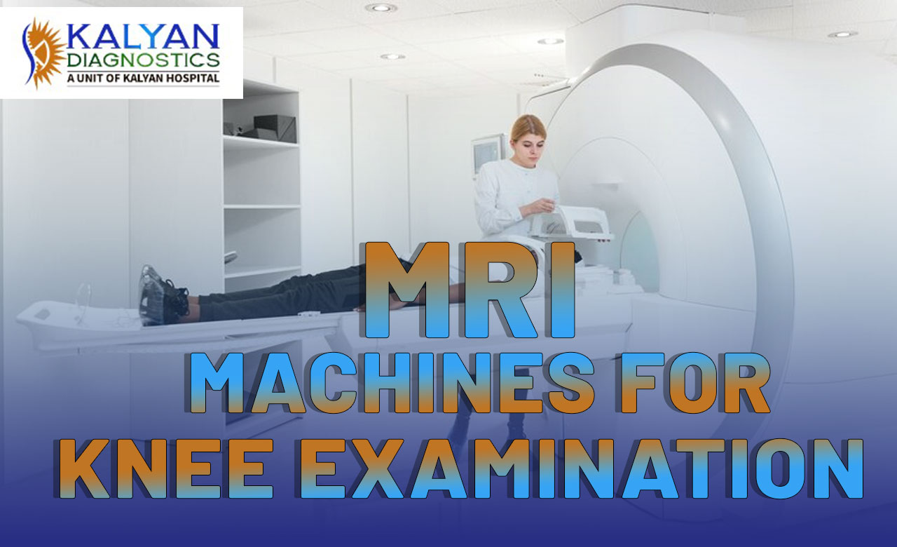
What can you expect during the knee MRI? 

You have to follow the following procedure during the MRI. 

  • Clothing: After reaching the hospital, you have to change your clothes and wear a gown for taking the X-rays.  
  • Remove Metal Items: Before the MRI, you’ll need to remove any metal objects, including jewelry, watches, and hairpins, as they can interfere with the magnetic field.


Procedure:

  • Positioning: You will normally be placed on your back on a movable examination table. A coil, a crucial component of the MRI scanner, will be used to hold the knee that is being photographed.
  • Securing: During the imaging procedure, you may be assisted in remaining still and maintaining the proper posture with the use of straps or cushions.
  • Contrast Agent: To make particular structures more visible, a contrast agent may occasionally be injected into a vein. An MRI of the knee is not always necessary.
  • Machine Noise: There may be loud tapping or knocking noises from the MRI scanner while the scan is being done. Earplugs or headphones may be offered to you to help you cope with the noise.
  • Communication: During the process, you will be able to speak with the MRI technologist, most often via an intercom system. As they lead you through the procedure, they will advise you when to keep still or shift positions.


Types of knee MRI machines. 

Mainly there are three types of MRI machines. 

  • Extremity MRI: This is a diagnostic imaging procedure that uses a closed MRI machine to look at the tissues in the arms and legs. 
  • Open MRI: Open MRI machines also stray from the traditional design of the full-body MRI, which makes some patients feel anxious due to the “closed-in” nature of the machine. An open MRI is open on the sides or features wider openings, still requiring the patient to lie on a sliding table.
  • 3 Tesla MRI: This type of closed MRI uses magnetic fields that have double the strength of a traditional MRI machine, thus producing an even more detailed image in less time.

The diagnostic centre does the MRI with a prescription that is given by a doctor who is well-qualified and experienced. Book an appointment with Kalyan Diagnostics for better-quality scans. It is also one of the best centers for MRI scans and a genuine place for affordable  MRI Cost in Ludhiana.

What is the difference between MRI and Contrast MRI?

Many people are suffering from different problems. Different types of tests are helpful to know about the different problems that can occur in your body. MRI is a type of scan that can create clear and detailed images of the situation of the organ inside the body. In this type of scan, magnetic and radio waves are used to create the images on the computer. It is also used for text neck diagnosis of cervical pain. To get a quality MRI, book an appointment with the best diagnostic centre in Ludhiana

What is the definition of MRI? 

MRI stands for Magnetic resonance imaging. The majority of people have heard about the MRI, which is an abbreviation. Radio Waves and powerful magnets are used to take the image from the depth of the human body. MRI images give you the correct and proper information about each and everything that is going on inside your body. An MRI report gives you appropriate information about your health conditions.  

What is the definition of Contrast MRI? 

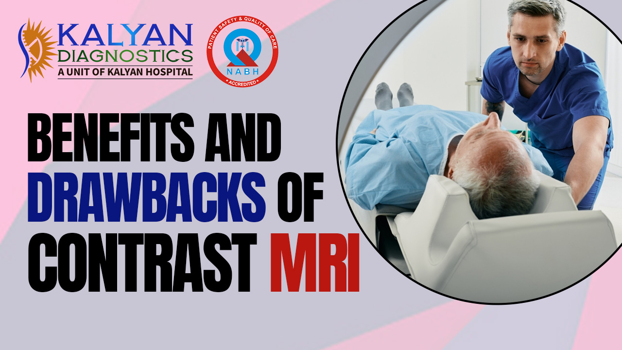
Contrast MRI is used to improve the visibility of internal human body structures in magnetic resonance imaging. The most common compound for contrast enhancement is gadolinium. The paramagnetic metal ion is commonly used in contrast to MRIs because of the way it travels within the magnetic fields.

What is the use of contrast MRI? 

The contrast MRI is used to Assess MS, cancer diagnosis and image clarity. 

  • Assessing MS: The greatest way to track the progression of multiple sclerosis is contrast MRI. MRI scans have transformed the radiologist’s diagnosis of MS, and it is the primary method that doctors use to diagnose the condition. Also, to the exclusion of other techniques like CT Scans and MRI scans, the lesions appear quite distinctly. 
  • Cancer diagnosis: Many doctors and radiologists use MRI dye during the MRI exam to make parts of your soft tissue look more detailed, they can spot tumors better as they start growing. If you have a cancer that’s just beginning to grow, it can be more challenging for the radiologist to spot.  
  • Image clarity: The MRI scan with contrast works with specific parts of the soft tissues, helping the radiologist diagnose your condition. In particular, tumor growth or other developments are more straightforward for the radiologist to identify when using contrast with the MRI scan.

What are the advantages of the Contrast MRI? 

Contrast MRI is beneficial in different ways. 

  • Enhanced nature: The radiologist uses contrast during the MRI, and the doctor generally requires additional scans because of the improved quality of scans. 
  • Measuring and evaluating tumors: A contrast MRI is superior at measuring and assessing tumors. Adding contrast makes it possible for the radiologist to detect even the smallest tumor and provides information about the precise location of the tumour.
  • Better Scan: The radiologist can interpret an MRI contrast scan better since they have more clarity and generate better-quality images.
  • Cost Effective: MRI is a type of scan that gives you an idea about internal health. MRI Cost in Ludhiana is only 3500-7000 bucks. 
The diagnostic center does the MRI with a prescription given by a well-qualified and experienced doctor. Book an appointment with Kalyan Diagnostics for better-quality scans. It is also one of the best centers for MRI scans.

What is Text neck Diagnosis of cervical pain of MRI?

Many people are suffering from different problems. Different types of tests are useful to know about the different problems that can occur in your body. MRI is a type of scan that can create clear and detailed images of the situation of the organ inside the body. In this type of scan, magnetic and radio waves are used to create the images on the computer. It is also used for text neck diagnosis of cervical pain. For getting a quality MRI, book an appointment with the best Diagnostic centre in Ludhiana

What is the definition of MRI? 

MRI stands for Magnetic resonance imaging. The majority of people have heard about the MRI, which is an abbreviation. Radio Waves and powerful magnets are used to take the image from the depth of the human body. MRI images give you the correct and proper information about each and everything that is going on inside your body. An MRI report gives you appropriate information about your health conditions. 

What is the definition of text neck syndrome? 

The text neck syndrome is also called tech neck. It is used to describe neck pain and damage stemming that is caused by looking down for a long time at mobiles, tablets and other handheld devices. Another reason for neck pain is poor posture and weak bones of the spine. Nowadays, people spend a lot of time on smartphones and computers, which increases the stress on the neck, and pain begins.

Cause of text neck syndrome

There are several factors that are responsible for text neck syndrome. 

  • Muscle strains: Spending more hours on smartphones and computers can cause muscle pain; even minor things like reading in bed can also affect the muscles. 
  • Worn joints: As with other joints in the body, neck joints tend to wear with age. In response to this wear and tear, the body often forms bone spurs that can affect joint motion and cause pain.
  • Nerve compression: Herniated disks or bone spurs in the vertebrae of the neck can press on the nerves branching out from the spinal cord.
  • Injuries: Rear-end auto collisions often result in whiplash injury. It occurs when the head jerks backwards and then forward, straining the soft tissues of the neck.
  • Diseases: Certain diseases, such as rheumatoid arthritis, meningitis or cancer, can cause neck pain.

Symptoms of text neck syndrome 

You will get several symptoms when you need an MRI Scan for the treatment of neck pain. 

  • Severe pain: Pain in the neck, shoulder or upper back is a typical characteristic of text neck. It can range from a dull ache to a sharp pain.
  • Muscle tightness and spasms: Your muscles of the neck become tight and sometimes swell. 
  • Decreased ability to move the head.
  • Reduce mobility: Text neck can cause tightness, making it difficult to move your neck.
  • Headache: Chronic headaches can stem from tight suboccipital muscles. Posture aside, excessive time spent looking at screens can also increase the risk of headaches and eyestrain.

Treatment of text neck syndrome with some preventions.

  • Rest and Avoiding Device Use: The first step in treating text neck injury is to rest the affected area and avoid the use of mobile devices, computers or any other activities that exacerbate the pain.
  • Ice or Heat Therapy: Applying ice or heat to the affected area can help reduce pain and inflammation. Ice therapy is recommended during the first 24-48 hours after injury, while heat therapy can be used after that to help relax the muscles.
  • Physical Therapy: Physical therapy can help relieve pain and stiffness and improve the range of motion by using exercises and stretches that focus on the neck and upper back muscles.
  • Massage Therapy: Massage therapy can help reduce muscle tension and increase blood flow to the affected area, which can help relieve pain and improve flexibility.
  • Posture Correction: Correcting poor posture habits can help prevent future text neck injuries. Simple changes like raising the device screen to eye level, taking frequent breaks, and stretching can make a big difference.

Cervical MRI for Neck pain 

An MRI Scan can be beneficial just to get the information and condition of your bones. An MRI Scan is a radiation-free and noninvasive imaging technique that provides you with concrete evidence about the suspected issues. In this procedure, powerful magnets, computer technology and detailed imaging techniques are used to click the images. 

Nowadays, before the treatment, doctors suggest an MRI Scan. For an MRI Test in Ludhiana, visit the best diagnosis center named Kalyan Diagnosis.                     

All You Need To Know About Dense Breast

Dense breasts, a condition where there is a higher proportion of glandular and connective tissue compared to fatty tissue, have become a topic of growing concern and interest in women’s health. Today, we aim to provide answers to commonly asked questions about dense breasts, shedding light in this often misunderstood aspect of breast health. 

Commonly Asked Questions

What are dense breasts?

Dense breasts refer to breasts with a higher amount of glandular and connective tissue than fatty tissue. This density can make it challenging to detect abnormalities on a mammogram since both cancerous and dense tissue appear white on the scan. 

Why do some women have dense breasts?

Factors, including genetic, age, and hormonal changes can lead to dense breasts. Younger women and those with a family history of dense breasts may be more prone to this condition.

Is having dense breasts normal?

Yes, having dense breasts is entirely normal and common. However, it’s essential to be aware of the potential challenges it poses for breast cancer detection.

Does having dense breasts increase breast cancer risks?

Dense breasts can make it more challenging to detect tumors on mammograms. Women with dense breasts may need additional screening methods, such as ultrasound or MRIs, to ensure accurate cancer detection.

How are Dense Breasts Identified?

Breast density is typically identified through a Mammography Test in Ludhiana. Radiologists use a grading system to categories ranging from predominantly fatty to extremely dense. 

Should I be concerned if I have dense breasts?

Having dense breasts doesn’t necessarily warrant concern, but it does highlight the importance of proactive breast health. Women with dense breasts should maintain regular screening and discuss additional imaging options with their healthcare provider. 

What additional screening options are available for women with dense breasts?

Women with dense breasts may benefit from supplementary screening, such as breast ultrasound or breast MRI. These methods can enhance the accuracy of cancer detection in dense breast tissue. 

Why does breast density matter?

Breast density matters because it can make it more challenging to detect abnormalities on a mammogram, dense breast tissue appears white on mammograms, similar to tumors and cysts, which can also appear white. This makes it harder to distinguish between normal tissue and potential health issues. 

Can dense breasts be inherited?

There is a genetic component to breast density, if your mother or sister has dense breasts, you may be more likely to have them as well. However, dense breasts can also occur in women with no family history or breast cancer.

Can lifestyle choices impact breast density?

Lifestyle choices like diet and exercise can influence overall breast health. However, maintaining a healthy lifestyle can contribute to overall well-being and reduce the risk of various health issues, including breast cancer. 

Conclusion

Understanding dense breasts is crucial for women’s health. If you have concerns or questions about your breast density, consult with the best healthcare professional at Kalyan Diagnostics Centre, a renowned Diagnostic center in Ludhiana, for personalized guidance and appropriate screening measures.

Understanding Dense Breasts: Evaluation and Treatment

Dense breasts have become a significant topic of discussion in women’s health, raising awareness about the importance of early detection and specialized care.

Dense breasts are a lesser-known aspect of women’s health that holds significant importance in breast cancer detection and prevention. Today, we will delve into the intricacies of dense breasts, their evaluation, and the pivotal role played by the mammography and Diagnostic Center in Ludhiana.

Understanding Dense Breasts

Dense breasts have a higher proportion of glandular and connective tissue compared to fatty tissue, this composition can make it challenging to detect abnormalities through traditional screening methods. It’s important for women to understand their breast density as it can influence the effectiveness of breast cancer screening.

Evaluation of Dense Breasts

Mammograms

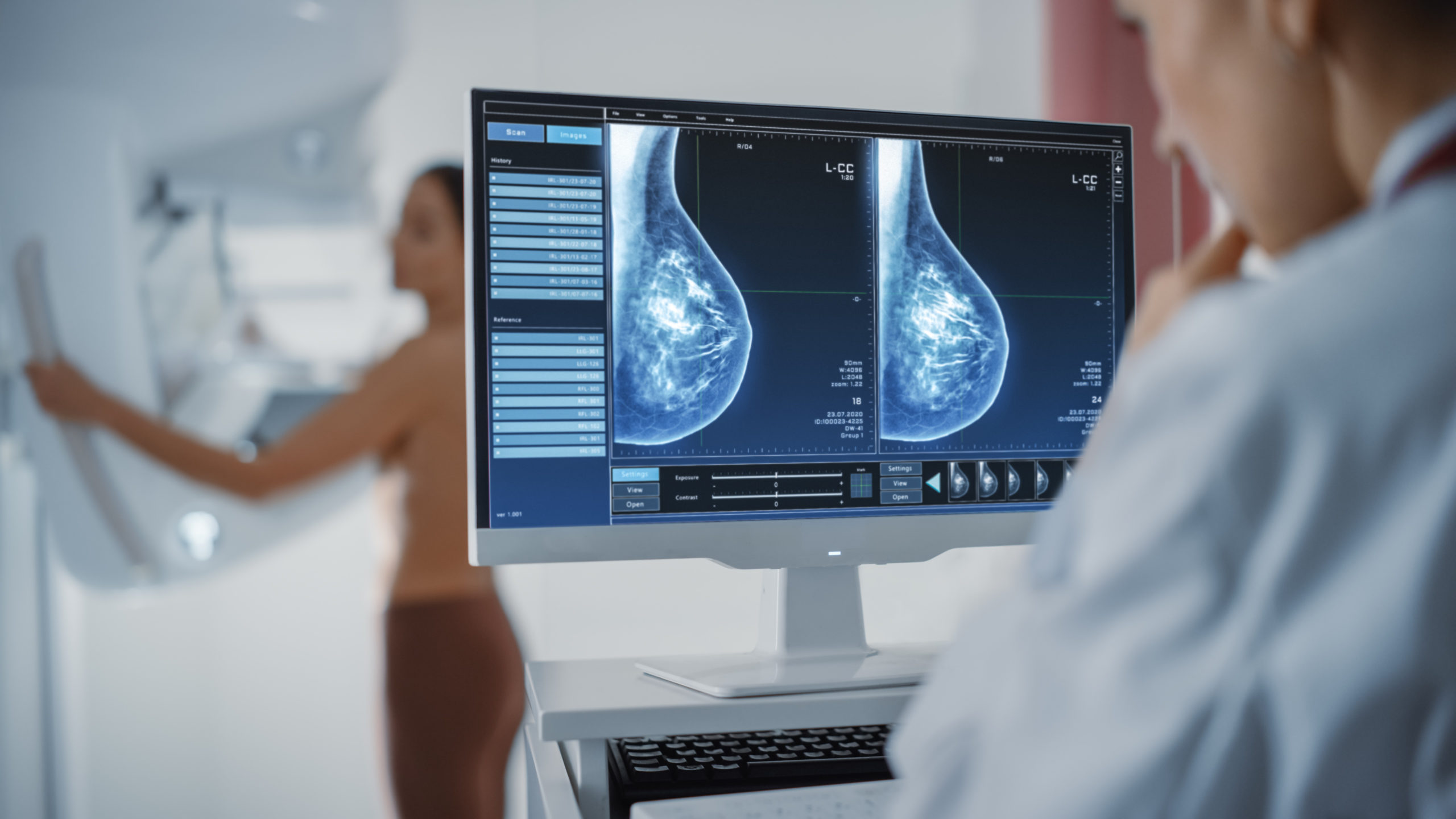
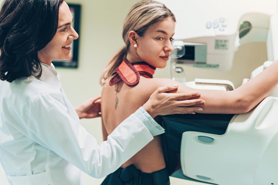
Mammography is a specialized imaging technique that uses low-dose X-rays to capture detailed images of breast tissue. It is a crucial tool for evaluating breast health, especially in cases of dense breast.

Women with dense breasts often undergo mammograms as part of their routine screenings. Mammography aids in detecting potential abnormalities, including tumors or calcifications that may be indicative to breast cancer.

Additional Imaging Modalities

To improve detection in dense breasts, additional imaging modalities may be recommended. This could be;

Ultrasound

Utilizing sound waves to create images, ultrasound is effective in identifying abnormalities that may be masked by dense tissue.

MRI

MRI provides detailed images of the breast and is especially useful in high-risk situations.

Treatment Options for Dense Breasts

While breast density is not something that can be altered, there are strategies for managing the associated challenges, particularly in the context of breast cancer screening and prevention.

1. Supplemental Screening

Women with dense breasts may benefit from supplemental screening methods in addition to mammography. This could involve regular ultrasound or MRIs, providing a more comprehensive view of breast tissue.

2. Genetic Testing

Understanding your genetic predisposition to breast cancer can be crucial. Genetic testing, especially for mutations in the genes, can help assess the risk and guide personalized screening and prevention strategies.

3. Lifestyle Changes

Maintaining a healthy lifestyle can contribute to overall breast health. These lifestyle choices may not directly alter breast density but can positively impact overall well-being and potentially reduce cancer risk.

Conclusion

Understanding and addressing dense breasts involve a comprehensive approach, from awareness and evaluation to advanced screening methods, Kalyan Diagnostics Centre plays a pivotal role in this journey, providing women with the necessary tools and expertise to prioritize their breast health schedule your Mammography Test in Ludhiana for a proactive step towards maintaining your well being.

Is Mammography Testing the Right Choice for You?

Welcome to our blog on diagnostic centers and mammography tests. If you have ever wondered about these essential healthcare services, you are in the right place. In simple terms, diagnostic centers are places where doctors use special machines to check your body’s health. Mammography tests, specifically for women, help detect breast problems. We’ll explain everything in a way that’s easy to understand so you’ll be well-informed and prepared for your health journey. If you are finding the right Diagnostic center in Ludhiana, it is crucial for your well-being.

Breast Cancer Screening: Thermogram No Substitute for Mammogram

Breast cancer is a significant health concern, and early detection is crucial. It’s essential to recognize that although thermograms may appear as an option for mammograms, they are not a replacement. Let’s break down why:

  • Different Approaches

Thermograms use heat to detect changes in breast tissue. They capture temperature variations, which can be linked to potential issues. Mammograms, on the other hand, rely on X-rays to create detailed images of the breast. This method can reveal even the tiniest abnormalities.

  • Reliability Matters

Mammograms have long been the gold standard for breast cancer screening. They are trusted and have a well-established track record. Thermograms, while intriguing, are less reliable when it comes to detecting small tumors. Relying solely on thermograms could lead to missed opportunities for early intervention.

If you’re concerned about breast health, consider heading to a diagnostic center for a mammography test. It’s a decision that could ultimately save your life.

Mammography vs. Thermography

When considering breast health, two frequently mentioned diagnostic tools are mammography and thermography. But what are they, and how do they differ?

Mammography is like a special X-ray for breasts. It uses low-dose radiation to create detailed images. It’s great at detecting small lumps or tumors, especially in older women. But the radiation can be a concern for some.

Thermography, on the other hand, is all about heat. It captures infrared images of your breasts to detect temperature changes. It’s radiation-free and safe, making it suitable for younger women. But it might not spot small lumps as effectively as mammography.

Each has its pros and cons, and your doctor will help you decide which is best for you. Remember, early detection is key for breast health.

Advice for Patients Getting Breast Cancer Screening

Breast cancer screening is vital for your well-being, and it’s not as daunting as it may seem. The process is painless, quick, and non-invasive. When preparing for your mammography test, remember these three essential points:

  • Choose a reputable Center:

Select a diagnostic center with a solid reputation and experienced staff. Research and read reviews to ensure you’re in capable hands. 

  • Communication is Key:

Don’t hesitate to voice any concerns or questions to the technicians. They are there to assist and make you comfortable throughout the process.

  • Prepare and Stay Positive:

Dress in comfortable clothing, free from deodorant or lotion, for accurate results. Regardless of the outcome, stay positive and seek support from loved ones. Remember, regular screenings are your best defense against breast cancer, promoting a healthier and happier you.

Conclusion

When it comes to your well-being, our center stands out as the ideal choice for a Mammography Test in Ludhiana. By opting for our services, you ensure accuracy, professionalism, and a commitment to your health. Regular screening at Kalyan Diagnostics Centre is your best ally for a healthier future.

The importance of mammographic tests to examine breast issues

Mammographic tests are beneficial for examining the different issues of breasts. The mammographic tests used by the doctors for analyzing the breast tissue. Mammography is an X-ray imaging method used to examine the breast for the early detection of cancer and other breast diseases. It is used as both a diagnostic and screening tool.

What is a mammographic test?

Mammography is an X-ray procedure. The X-rays taken from a mammographic machine are known as mammograms. These mammograms are useful for a doctor to detect breast cancer at an early level. Regular mammograms can identify breast cancer. 

Reason for the mammograms 

A mammogram is a type of X-ray picture of the breast. Mammograms are helpful to examine the early signs of cancer of the breast. Daily basis mammograms are lucrative for finding breast cancer early. The human body takes a minimum of three years to notice the symptoms of breast cancer. The Mammography Test in Ludhiana is done at Kalyan Diagnostics.

Symptoms of breast cancer

People notice many symptoms if they are suffering from breast cancer. You may notice the following symptoms. 

  • You may feel lumps in the area of the breast and underarm. 
  • Feel swelling and thickening of the breast. 
  • Feel irritation and dimple breast skin. 
  • Redness and breaking skin of the nipple area and area of the breast.
  • Pain in any area of the breast.
  • Pain around the nipple area. 
  • Discharge from the nipple other than the milk, blood also includes.  
  • Notice the change in shape and size of the breast. 

If you notice any symptoms, then you should go to the best Diagnostic center in Ludhiana. Kalyan Diagnostics is considered the best diagnostic center. 

Cause of breast cancer 

Many things are responsible for breast cancer. Things such as age, hormonal medications, contraceptive pills and lifestyle factors can cause breast cancer. 

Natural factors

  • Age: Breast cancer occurs after the age of 50. The menstrual cycle stops after 50. Menopause is a reason for breast cancer after the age of 50.
  • Background history: if anyone in your family has ovarian cancer, it is at risk of breast cancer. There are the chances that you will also suffer from breast cancer because cells of cancer are also present in the genetic material. 
  • The dense tissue: The woman’s breast contains thousands of tiny glands. These are called lobules. Lobules are also known as milk glands. The tissue of glands is very dense. Women who have denser tissue are at the risk of breast cancer because it gives more space for the generation of cancer cells.

Hormones and hormonal medicines:  

  • Estrogen: An important hormone secretion named estrogen also causes breast cancer. The storage for eggs, called ovaries, starts producing estrogen at the start of puberty. It helps to regulate your periods. 
  • HRP: Hormonal replacement therapies can cause breast cancer if it is taken more than once a year. Except for vaginal estrogen, other HRP can be a reason for breast cancer. 
  • Contraceptive pills: Many women take contraceptive pills. The studies of the researcher show that the pills have a component that increases the risk of breast cancer. 

Lifestyle of a person 

  • Alcohol and drugs: Alcohol and drugs can increase the risk of breast cancer. 
  • Increase in weight: Obesity is a major problem. Many problems occur because of obesity. Obesity can be responsible for a high risk of breast cancer.
  • Radiation: People who are working in laboratories where X-ray and CT scans are done are at risk of breast cancer. These radiations are really dangerous for health. If you had an X-ray and CT done, then the chances of breast cancer are there. 

Diagnosis for breast cancer.

Doctors suggest you take different kinds of X-rays. With the help of these X-rays, doctors decide the treatment plan. 

  • Physical examination of breast
  • X-ray
  • Mammograms
  • Magnetic Resonance Imaging. 
  • CT scan 
  • Sample of cells of the breast
  • Ultrasound of breast

Treatment of breast cancer

There are two types of treatment according to the level of breast cancer: surgery and therapies. 

Surgery treatment

Surgeries are helpful in removing the lumps and cancer cells from the breast. The steps are as follows:

  • Lumpectomy: In this step, only cancer cells are removed
  • Mastectomy: Mastectomy is to remove the breast. It mostly performs in highly affected patients. 
  • Sentinel node biopsy: in this surgery, a limited number of lymph nodes are removed. 
  • Axillary lymph node dissection: Many lymph nodes are removed in this.
  • Removal of both breasts: Many women have breast cancer on one side only. They have to choose the removal of another breast because of genetic predisposition. 

Types of Therapies

Several types of therapies are beneficial for reducing the risk of breast cancer.

  • Radiation therapy
  • Chemotherapy
  • Hormonal therapy 
  • Immunotherapy

Breast cancer is a common problem in women in today’s era. The reason is an unhealthy diet and a busy lifestyle. To reduce the chances of breast cancer, females should eat the proper diet and live a healthy lifestyle.