What Doctors Seek in Your Mammogram Results


What Doctors Seek in Your Mammogram Results

A radiologist is the person who checks the results of your mammography. Radiologists of the best Diagnostic center in Ludhiana use x-ray images to find out about any hidden illness or problems in your skin.  

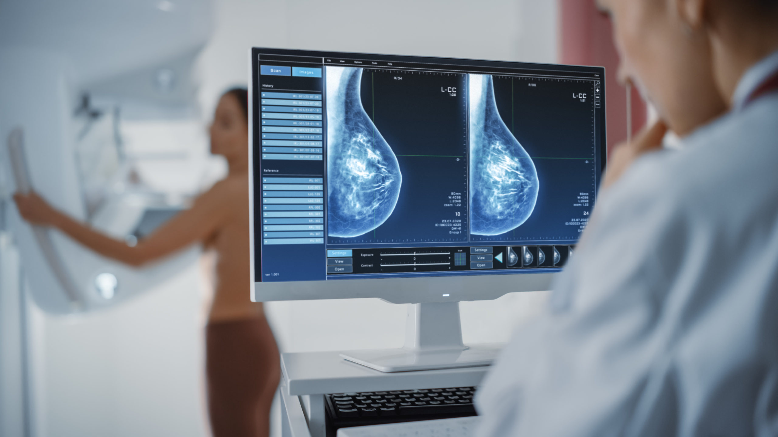
While seeing your mammogram reports, doctors analyze them with your old mammogram reports. This helps the radiologist to find out if there is anything new on your mammogram this time or not. If new mammogram reports are similar to the old ones, that means no further tests are needed. 

Radiologist searches for the various kinds of changes in your breasts by mammography, like white spots of calcification, strange masses area, and other symptoms that possibly can be the symptoms of cancer. 

Calcification 

There are mainly two types of calcifications. They may or may not be symptoms of cancer. Cancer signs show as white spots on mammography reports. 

Macrocalcifications

Microcalcification is the result of the deposition of calcium tissues. Which often occurs due to old injuries and aging. In general, these are not related to cancer, and biopsy-like additional treatments are not needed. 

It is common in women and starts deposition after the age of 50. 

Microcalcifications

Microcalcifications refer to the small calcium element deposition in the breasts. Microcalcifications are a matter of concern, but they may not be the symptoms of cancer. These tiny calcium deposits help doctors to find out about the possibility of cancers. 

Biopsy is not needed for microcalcification. But if doctors find any strange pattern in them, then experts will suggest a biopsy. 

Masses

There is a special spot on the breasts for the mass, which looks different from other areas of the breasts. Masses can have different possibilities, such as cysts and noncancer hard tumors. Undoubtedly, it can be a symptom of cancer, too. If anybody notices masses in their breasts, then a Mammography Test in Ludhiana at Kalyan Diagnosis is necessary. 

Suppose the doctor is not sure about the lump. Whether it is a fluid-filled cyst or a solid mass, then they might use a needle to see what’s inside the lump. After fluid removal, if a lump disappears, then it might be the just lump, and no further tests are needed. 

But if a lump has no fluid inside, then it is a matter of cancer, and further tests are necessary. 

Asymmetry 

Asymmetry on mammogram reports resembles white spots, which is not normal and similar to breast tissue patterns. It had types like focal asymmetry and global asymmetry. 

Asymmetric patterns are not an indication of cancer, but further tests are necessary to find out whether you have cancer or not. 

Breast Density

Mammogram reports tell about breast density. This tells about the shape and firmness of your breast along with fatty tissues in it. If your breasts are labeled as dense, that means your breasts have more fibrous and glandular tissue instead of fatty tissues. 

एमआरआई (MRI) सर्वाइकल स्पाइन करवाने के क्या है लागत, परिणाम व सम्पूर्ण जानकारी ?

एमआरआई (MRI) जोकि हर बीमारी को पकड़ने में काफी मददगार साबित होती है। साथ ही एमआरआई की मदद से हम अंधरुनि समस्या को जानकर उसका इलाज अच्छे से करवा सकते है। वहीं एमआरआई से सर्वाइकल स्पाइन का इलाज व जाँच को कैसे किया जाता है, इसके बारे में आज के लेख में चर्चा करेंगे ;

एमआरआई (MRI) कैसे सहायक है सर्वाइकल स्पाइन की जाँच के लिए ?

  • एमआरआई सर्वाइकल स्पाइन रीढ़ के सर्वाइकल क्षेत्र (गर्दन के पीछे की तरफ) की जांच और पता लगाने में मदद करता है। यह गर्दन और रीढ़ की अंदरूनी चोट या फ्रैक्चर का पता करने में मदद करते है। 
  • यदि आपको गर्दन के क्षेत्र में दर्द है तो डॉक्टर आमतौर पर इसकी सलाह देते है। यह परीक्षण गर्दन और सर्वाइकल स्पाइन के कोमल ऊतकों का 3-डी दृश्य देता है। वही लुधियाना में MRI का खर्च या सर्वाइकल स्पाइन की कीमत 3000 रुपये से लेकर 6000 रुपये तक है।

सर्वाइकल स्पाइन के MRI में क्या शामिल हो सकते है ?

  • सात कशेरुक जो C1 से C7 तक होते है। 
  • रक्त वाहिकाएं। 
  • नसे। 
  • पट्टा। 
  • बंधन। 
  • उपास्थि आदि इसके MRI में शामिल होते है। 

क्या है सर्वाइकल MRI स्कैन ? 

  • सर्वाइकल एमआरआई (मैग्नेटिक रेजोनेंस इमेजिंग) गर्दन के पीछे की हड्डियों, जिन्हें सर्वाइकल स्पाइन कहा जाता है, की विस्तृत छवियां उत्पन्न करने के लिए एक चुंबकीय क्षेत्र और रेडियो तरंगों का उपयोग करते है।  
  • परीक्षण के दौरान, एमआरआई मशीन व्यक्ति के शरीर में एक अस्थायी चुंबकीय क्षेत्र बनाती है, जो शरीर में मौजूद हाइड्रोजन परमाणुओं को क्षेत्र के साथ संरेखित करने के लिए मजबूर करती है।
  • अगर आप गर्दन के पीछे की हड्डियों या सर्वाइकल स्पाइन की समस्या से परेशान है, तो इससे बचाव के लिए आपको लुधियाना में बेस्ट रेडियोलॉजिस्ट से अपनी जाँच को करवाना चाहिए।

सर्वाइकल स्पाइन MRI का उद्देश्य क्या है ?

  • रीढ़ की हड्डी में चोट लगी हो तो इसके लिए इस स्कैन का चयन किया जाता है।  
  • हड्डियों या कोमल ऊतकों में ट्यूमर या कैंसर होने पर भी आप इस स्कैन का चयन कर सकते है। 
  • हर्नियेटेड डिस्क होने पर इस स्कैन का चयन करना।
  • डिस्क फलाव की समस्या होने पर इसका चयन करना। 
  • जन्मजात या शारीरिक जन्म दोष होने पर इस स्कैन का चयन करना।
  • हड्डियों की बनावट में असामान्यता के आने पर इसका चयन करना।
  • स्पाइनल स्टेनोसिस की समस्या होने पर इसका चयन करना। 
  • ऊपरी अंगों में अकड़न होने पर इसका चयन करना।

MRI सर्वाइकल स्पाइन के विभिन प्रकार क्या है ?

  • WSS के साथ MRI का सर्वाइकल स्पाइन करवाना। 
  • एमआरआई सर्वाइकल स्पाइन फ्लेक्सन-एक्सटेंशन। 
  • ब्रेन जांच के साथ एमआरआई सर्वाइकल स्पाइन की जाँच। 
  • मल्टीपल स्केलेरोसिस का पता लगाने के लिए। 
  • ALS के पहचान के लिए। 
  • गतिशील एमआरआई सर्वाइकल स्पाइन।
  • कंट्रास्ट के साथ एमआरआई सर्वाइकल स्पाइन। 

सर्वाइकल स्पाइन MRI क्यों किया जाता है ?

  • गर्दन, बांह या कंधे में गंभीर दर्द जो इलाज के बाद भी कम ना हो तो इस जाँच का चयन करें। 
  • पैर में कमजोरी या सुन्नता जैसे लक्षणों के साथ गर्दन में दर्द होने पर। 
  • रीढ़ की हड्डी के जन्म दोष या रीढ़ की हड्डी में चोट का पता लगाना। 
  • रीढ़ की हड्डी में संक्रमण या ट्यूमर का पता लगाने के लिए। 
  • रीढ़ की हड्डी में गठिया का पता लगाने के लिए। 
  • परीक्षण स्पाइन सर्जरी से पहले या बाद में भी किया जा सकता है।

सर्वाइकल MRI कैसे किया जाता है ?

  • रेडियोग्राफर आपको स्कैन कक्ष में प्रवेश करने से पहले डेन्चर और विग सहित सभी धातु की वस्तुओं को हटाने का निर्देश देंगे। 
  • वे आपको हॉस्पिटल गाउन पहनने के लिए कह सकते है। 
  • आपको एक बिस्तर पर लेटना होगा, जो एमआरआई स्कैनर मशीन के अंदर चला जाएगा। 
  • प्रक्रिया में एक डाई (कंट्रास्ट) की आवश्यकता हो सकती है, जो परीक्षण से पहले आपकी बांह में एक नस के माध्यम से दी जाएगी। 
  • कंट्रास्ट कुछ क्षेत्रों को अधिक साफ-साफ देखने में मदद करता है। 
  • आपको बिल्कुल स्थिर होकर लेटना होगा, क्योंकि हरकतें तस्वीरों को धुंधला बना सकती है। 
  • परीक्षण आमतौर पर 30-60 मिनट तक चलता है लेकिन इसमें अधिक समय भी लग सकता है। 

सर्वाइकल MRI के जोखिम क्या है ?

  • कंट्रास्ट से एलर्जी की प्रतिक्रिया को करना, जोकि काफी जोखिम भरा होता है। 
  • एमआरआई मशीन में मौजूद मजबूत चुंबक पेसमेकर और अन्य धातु प्रत्यारोपण में खराबी या शरीर के अंदर हिलने-डुलने का कारण बन सकते है। 
  • उपयोग किया जाने वाला कंट्रास्ट उन किडनी रोगों वाले लोगों के लिए हानिकारक हो सकता है जिन्हें डायलिसिस की आवश्यकता होती है।

सुझाव :

सर्वाइकल स्पाइन में अगर आपको दिक्कत है तो इस समस्या का सामना करने के लिए आपको एमआरआई का चयन करना चाहिए, क्युकी एमआरआई की मदद से ये जानना काफी आसान हो जाता है की व्यक्ति को किस तरह की समस्या है। 

इसके अलावा आप चाहें तो इस टेस्ट को कल्याण डायग्नोस्टिक्स हॉस्पिटल से भी करवा सकते है। 

एमआरआई (MRI) स्कैन स्पाइन लम्बर यानि (पीठ में होने वाले दर्द) के मामलों में कैसे है सहायक ?

एमआरआई (MRI) स्कैन जोकि हर तरह की समस्या का स्पष्ट रूप में मूल्यांकन करते है साथ ही इसका चयन करके हमें सब कुछ स्पष्ट हो जाता है की हम किस तरह की बीमारी से जूझ रहें है और साथ ही एमआरआई (MRI) स्कैन डॉक्टरों के लिए काफी सहायक माना जाता है। इसके अलावा एमआरआई स्पाइन लम्बर की समस्या को कैसे विस्तार से बाहर लाने में हमारी मदद करेंगे इसके बारे में हम सम्पूर्ण लेख में चर्चा करेंगे ;

क्या है एमआरआई (MRI) स्कैन ?

  • MRI (मैग्नेटिक रेसोनेंस इमेजिंग) एक जांच की प्रक्रिया है। इसमें शरीर के अंदर की उच्च गुणवत्ता वाली, विस्तृत तस्वीरें बनाने के लिए एक बड़े चुंबक, रेडियो तरंगों और कंप्यूटर का उपयोग किया जाता है। 
  • एमआरआई जांच का चयन अगर कोई भी करता है, तो उसे दर्द की समस्या का सामना नहीं करना पड़ता, इसलिए इस स्कैन का चयन बच्चे भी कर सकते है। 

इस स्कैन का चयन करने से बेशक आपको दर्द की समस्या का सामना नहीं करना पड़ता, लेकिन जरूरी है की अगर आप इस स्कैन का चयन करने जा रहें है तो इससे पहले लुधियाना में MRI का खर्च कितना है इसके बारे में जरूर जानकारी हासिल करें।

एमआरआई (MRI) स्कैन से क्या-क्या बाते सामने आती है ?

  • एमआरआई स्कैन का चयन जब आप करते है तो आपको मस्तिष्क, रीढ़, जोड़ों, हृदय, लीवर, और कई अन्य अंगों जैसे शरीर के कोमल-ऊतक व अन्य संरचनाओं का पता एमआरआई (MRI) चित्रों के द्वारा आसानी से पता लग जाता है। 
  • कैंसर, हृदय की बीमारी, मांसपेशियों और हड्डियों की बीमारियों, और कई अन्य बीमारियों का डायग्नोसिस एमआरआई स्कैन से किया जा सकता है। इस स्कैन की मदद से हम रीढ़ की हड्डी व पीठ की हड्डी में दर्द की समस्या का भी पता लगा सकते है।

एमआरआई (MRI) स्पाइन लम्बर स्कैन में किस तरह की तकनीक का प्रयोग किया जाता है 

  • चुंबकीय अनुनाद इमेजिंग (एमआरआई) स्कैन एक सामान्य प्रक्रिया है, जिसका उपयोग रीढ़ की हड्डी और अन्य अंगों के निदान के लिए भी किया जाता है। 
  • इस प्रक्रिया में चुंबकीय तरंगों का इस्तेमाल रीढ़ की हड्डी के विस्तृत चित्र का निर्माण करने के लिए उपयोग किया जाता है। 
  • इसमें मरीज को मेज़ पर लेटा कर एमआरआई मशीन के अंदर ले जाया जाता है, जो, एक बड़े डोनट की तरह लगता है। 
  • एमआरआई स्कैन आक्रामक नहीं है और यह एक दर्द रहित प्रक्रिया है। 
  • वहीं एक्स-रे और सीटी स्कैन के विपरीत, एमआरआई स्कैन में विकिरण का उपयोग नहीं होता है, इसलिए यह एक सुरक्षित प्रक्रिया है।

एमआरआई (MRI) स्पाइन लम्बर के प्रकार क्या है ?

  • एमआरआई स्पाइन लम्बर के लिए कंट्रास्ट एजेंट शरीर में इंजेक्शन या मौखिक रूप से दिया जाता है। कंट्रास्ट एजेंट आमतौर पर बेहतर छवियाँ देता है, जबकि बिना कंट्रास्ट इसके विपरीत है। हालांकि, आपके डॉक्टर यह फैसला करते है कि आपको कौन-से टेस्ट के लिए जाना है।
  • अधिकांश एमआरआई स्कैन स्पाइन लम्बर में रोगी को लिटाकर मशीन में ले जाने की आवश्यकता होती है। जो लोग बंद मशीन में जाने से डरते है, उनके लिए यह एक चिंता का विषय हो सकता है। तकनीशियन उन मामलों में एनिस्थिसिया दे सकते है या वह ओपन एमआरआई का विकल्प भी सामने वाले मरीज को दे सकते है, पर ध्यान रहें ये स्कैन थोड़ा मेहगा है।
  • टेस्ला चुंबकीय शक्ति की माप की एक इकाई है। एमआरआई मशीन 1.5 टेस्ला या 3 टेस्ला हो सकती है। 3 टेस्ला एमआरआई स्कैन मशीन तेज गति से बेहतर गुणवत्ता की छवियों का उत्पादन कर सकती है। हालांकि, यह अधिक महंगी होती है। इसके अतिरिक्त, भारत में हो रहे अधिकांश एमआरआई स्कैन के लिए 1.5 टेस्ला मशीन पर्याप्त है।
  • स्पाइन लम्बर क्षेत्र के लिए आवश्यक जानकारी के आधार पर, डॉक्टर स्क्रीनिंग या सामान्य एमआरआई की सिफारिश कर सकते है। 

ध्यान रहें अगर आपको स्पाइन लम्बर करवाना है, तो इसके लिए आप लुधियाना में बेस्ट रेडियोलॉजिस्ट का चयन करें।

स्पाइन लम्बर स्कैन को क्यों कराया जाता है ?

  • पीठ के निचले हिस्से में लगातार दर्द अगर हो तो आप इस स्कैन का चयन कर सकते है। 
  • रीढ़ की हड्डी के निचले भाग में चोट लगने पर। 
  • मस्तिष्क या रीढ़ की हड्डी में कैंसर के लक्षण का पता लगाने पर। 
  • रीढ़ की हड्डी में जन्म से कोई समस्या होने पर। 
  • पैरों के सुन्न पड़ने पर। 
  • पीठ के निचले हिस्से में सर्जरी होने पर।

सुझाव :

अगर आप पीठ या रीढ़ की हड्डी में दर्द की समस्या से परेशान है तो इससे बचाव के लिए आपको स्पाइन लम्बर स्कैन को जरूर करवाना चाहिए, वहीं आप चाहें तो इस स्कैन को कल्याण डायग्नोस्टिक्स हॉस्पिटल से भी करवा सकते है।

लिवर फाइब्रोस्कैन उपचार क्या है और ये किस काम के लिए प्रयोग में लाया जाता है ?

लिवर फाइब्रोस्कैन एक नवीन चिकित्सा प्रक्रिया है जिसका उपयोग लिवर के स्वास्थ्य का आकलन करने में किया जाता है। यह लीवर की कठोरता को मापता है, जो फाइब्रोसिस या घाव का एक प्रमुख संकेतक है, जो विभिन्न लीवर स्थितियों के परिणामस्वरूप हो सकता है। यह गैर-इनवेसिव तकनीक लिवर फाइब्रोसिस के स्तर को निर्धारित करने के लिए एक विशेष अल्ट्रासाउंड मशीन का उपयोग भी करती है, तो आइये जानते है फाइब्रोसिस के बारे में ;

लिवर फाइब्रोस्कैन कैसे काम करता है ?

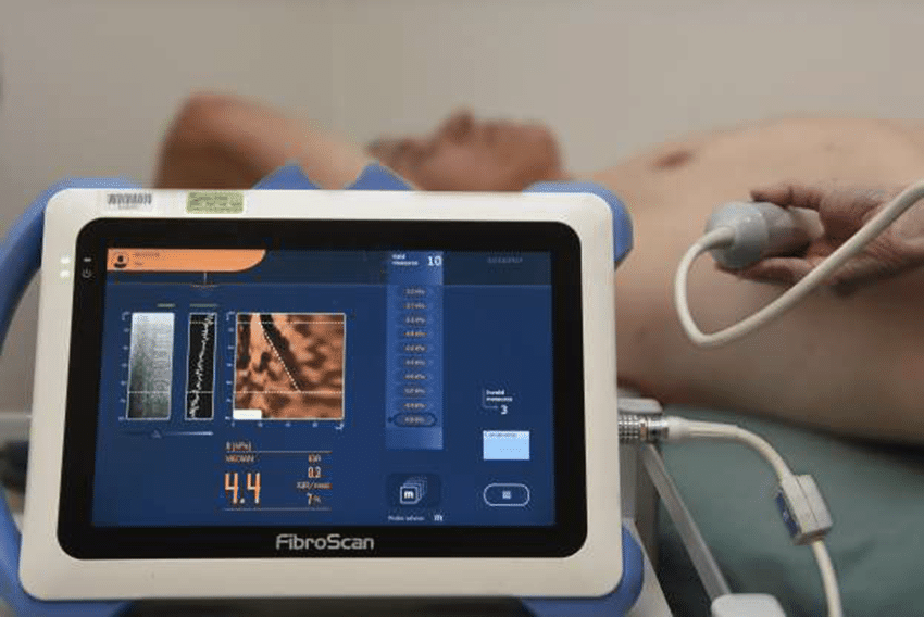
फाइब्रोस्कैन के दौरान, रोगी अपनी पीठ के बल लेट जाता है और तकनीशियन त्वचा की सतह पर, आमतौर पर पसली के दाहिने हिस्से पर जांच करता है। जांच त्वचा के माध्यम से यकृत तक हल्के कंपन उत्सर्जित करती है, जिससे कतरनी तरंगें बनती है, जिन्हें यकृत की कठोरता निर्धारित करने के लिए मापा जाता है। यह कठोरता रीडिंग लीवर के स्वास्थ्य का एक विश्वसनीय माप है। पूरी प्रक्रिया त्वरित, दर्द रहित है और इसमें एनेस्थीसिया की आवश्यकता नहीं होती है, जिससे यह कई रोगियों के लिए पसंदीदा विकल्प बन जाता है।

लिवर फाइब्रोस्कैन का उपयोग कैसे किया जाता है ?

  • लिवर फाइब्रोस्कैन का उपयोग मुख्य रूप से लिवर रोगों के निदान और निगरानी में किया जाता है, विशेष रूप से विभिन्न स्थितियों, जैसे हेपेटाइटिस सी, फैटी लिवर रोग, अल्कोहलिक लिवर रोग और अन्य के कारण होने वाले लिवर फाइब्रोसिस की प्रगति का आकलन करने में। फ़ाइब्रोस्कैन से प्राप्त परिणाम लीवर की क्षति की सीमा निर्धारित करने और उपचार संबंधी निर्णय लेने में सहायता करते है।
  • इसके अतिरिक्त, फ़ाइब्रोस्कैन परिणाम विशिष्ट हस्तक्षेपों की आवश्यकता का आकलन करने में मदद करते है, जैसे हेपेटाइटिस सी के लिए एंटीवायरल थेरेपी या फैटी लीवर रोग जैसी स्थितियों के लिए जीवनशैली में संशोधन। यह स्वास्थ्य देखभाल पेशेवरों को उपचार की प्रभावशीलता की निगरानी करने और आवश्यकतानुसार उन्हें समायोजित करने की अनुमति देता है, जिससे रोगी देखभाल के लिए अधिक व्यक्तिगत दृष्टिकोण की पेशकश की जाती है।

अगर आप अपने लिवर की जाँच को करवाना चाहते है तो इसके लिए आपको लुधियाना में फाइब्रोस्कैन का उपयोग जरूर से करना चाहिए।

लिवर फाइब्रोस्कैन के लाभ क्या है ?

  • लिवर फ़ाइब्रोस्कैन का मुख्य लाभ इसकी गैर-आक्रामक प्रकृति में शामिल है। परंपरागत रूप से, लीवर स्वास्थ्य मूल्यांकन में लीवर बायोप्सी जैसी आक्रामक प्रक्रियाएं शामिल होती है, जो न केवल असुविधाजनक होती है बल्कि कुछ जोखिम भी उठाती है। फ़ाइब्रोस्कैन इन जोखिमों और असुविधाओं को दूर करता है, जिससे यह एक सुरक्षित और अधिक रोगी-अनुकूल विकल्प बन जाता है।
  • इसके अलावा, यह जल्दी परिणाम प्रदान करता है, जिससे चिकित्सा पेशेवरों को रोगी के यकृत स्वास्थ्य का तुरंत मूल्यांकन करने और उपचार योजनाओं के बारे में समय पर, सूचित निर्णय लेने की अनुमति मिलती है। फ़ाइब्रोस्कैन की सहजता और दक्षता नियमित निगरानी को प्रोत्साहित करती है, जिससे बेहतर रोग प्रबंधन में योगदान मिलता है।

महत्वपूर्ण बातें !

लिवर फाइब्रोस्कैन तकनीक का विकास और व्यापक उपयोग लिवर रोग निदान और प्रबंधन में एक महत्वपूर्ण प्रगति का प्रतिनिधित्व करते है। इस प्रक्रिया की गैर-आक्रामक, तीव्र और सटीक प्रकृति वैश्विक स्तर पर लीवर की स्थितियों के निदान और प्रबंधन के तरीके को बदलने की क्षमता रखती है।

जैसे-जैसे लिवर स्वास्थ्य के क्षेत्र में अनुसंधान और प्रगति जारी है, लिवर फाइब्रोस्कैन के और अधिक विकसित होने की उम्मीद है, जो संभावित रूप से लिवर की स्थितियों में अधिक विस्तृत जानकारी प्रदान करेगा और इसके अनुप्रयोगों का विस्तार करेगा।

फाइब्रोस्कैन का उपयोग क्यों किया जाता है ?

  • फाइब्रोस्कैन एक गैर-इनवेसिव डायग्नोस्टिक टूल है, जिसका उपयोग लिवर के स्वास्थ्य का आकलन करने के लिए किया जाता है। यह लिवर की कठोरता को मापने के लिए एक विशेष प्रकार की अल्ट्रासाउंड तकनीक का उपयोग करता है जिसे क्षणिक इलास्टोग्राफी के रूप में जाना जाता है। फाइब्रोस्कैन का प्राथमिक उद्देश्य लिवर फाइब्रोसिस का निदान करना है, जो एक ऐसी स्थिति है जहां लिवर के ऊतक क्षतिग्रस्त हो जाते है और खराब हो जाते है, जिससे लिवर की शिथिलता हो जाती है।
  • लीवर फाइब्रोसिस कई कारकों के कारण हो सकता है, जिनमें हेपेटाइटिस बी और सी, शराब का दुरुपयोग और गैर-अल्कोहल फैटी लीवर रोग शामिल है। फाइब्रोस्कैन का उपयोग इन स्थितियों के कारण लीवर की क्षति की सीमा का पता लगाने के लिए किया जाता है।
  • प्रक्रिया दर्द रहित और गैर-इनवेसिव है, जो इसे लीवर बायोप्सी जैसी अधिक आक्रामक नैदानिक ​​प्रक्रियाओं का एक लोकप्रिय विकल्प बनाती है। फाइब्रोस्कैन के परिणाम डॉक्टरों को अपने रोगियों के लिए उपचार का सर्वोत्तम तरीका निर्धारित करने में मदद कर सकते है, जिसमें लिवर की कार्यक्षमता में सुधार के लिए दवा या जीवन शैली में बदलाव शामिल हो सकते है।
  • फाइब्रोस्कैन का उपयोग विभिन्न कारकों के कारण होने वाले लिवर फाइब्रोसिस के निदान के लिए किया जाता है और डॉक्टरों को उपचार का सर्वोत्तम तरीका निर्धारित करने में मदद करता है।

लुधियाना में फाइब्रोस्कैन का खर्च कितना आता है, इसके बारे में जानने के बाद ही इसका चयन करें।

याद रखें !

यदि आप लिवर की समस्या का सामना कर रहें है, तो इससे बचाव के लिए आपको कल्याण डायग्नोस्टिक्स सेंटर का चयन करना चाहिए।

निष्कर्ष :

लिवर फाइब्रोस्कैन एक अभूतपूर्व तकनीक है, जो लिवर स्वास्थ्य के मूल्यांकन और निगरानी में क्रांतिकारी बदलाव लाती है। इसकी गैर-आक्रामक प्रकृति, गति और सटीकता इसे यकृत रोगों के निदान और उचित उपचार रणनीतियों का निर्धारण करने में एक अमूल्य उपकरण बनाती है। अपनी निरंतर प्रगति के साथ, फ़ाइब्रोस्कैन रोगी की देखभाल बढ़ाने और यकृत की स्थिति वाले व्यक्तियों के परिणामों में सुधार करने में महत्वपूर्ण भूमिका निभाने के लिए तैयार है।

लिवर फाइब्रोस्कैन एक सरल, दर्द रहित और प्रभावी प्रक्रिया है। लिवर स्वास्थ्य के क्षेत्र में आशा की किरण के रूप में खड़ी है, जो दुनिया भर के रोगियों के लिए लिवर रोगों के बेहतर, अधिक कुशल प्रबंधन का वादा करती है।

How Mammograms Are Helpful For A Doctor

Mammograms are a scan that shows the deep structure of your breast. Many males and females are struggling with breast cancer. With an unhealthy diet and lifestyle, people are suffering from several problems. Mammographic tests give you various benefits. The mammographic tests used by the doctors for analyzing the breast tissue. Mammography is an X-ray imaging method used to examine the breast for the early detection of cancer and other breast diseases. It is used as both a diagnostic and screening tool.

What is a Mammographic test? 

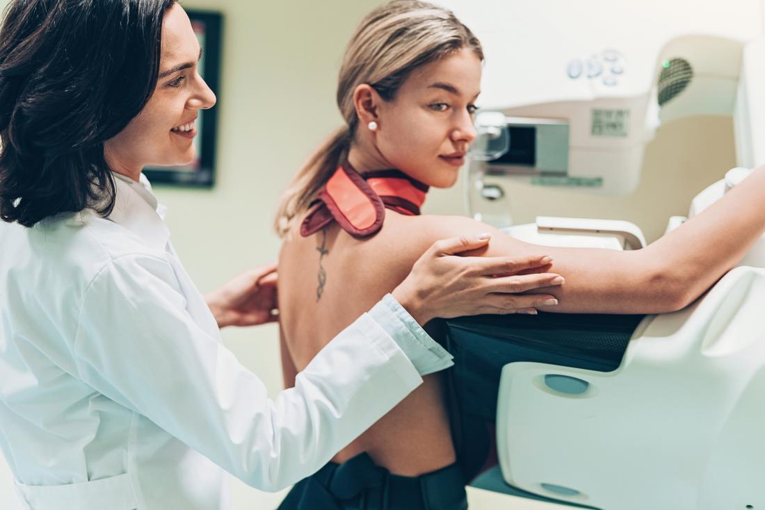
Mammographic tests are a type of X-ray that doctors use to look for the reason for breast cancer. Regular checkups of the breast can find the cancer of the breast at the initial stage. The X-rays taken from a mammographic machine are known as mammograms.

Reason for mammography test. 

A mammogram is a type of X-ray picture of the breast. Mammograms are helpful to examine the early signs of cancer of the breast. Daily basis mammograms are lucrative for finding breast cancer early. The human body takes a minimum of three years to notice the symptoms of breast 

cancer. The Mammography Test in Ludhiana is done at Kalyan Diagnostics.

Cause of breast cancer. 

Different factors are responsible for breast cancer. Breast cancer occurs when breast cells start to grow abnormally. The abnormal cells divide at a higher speed than normal cells. The division of abnormal cells causes lumps and mass under the skin of the breast. Cells spread to the Lymph nodes and the other parts of your body. Breast cancer begins in the milky ducts. Breast cancer also starts in the glandular tissue. The glandular tissue is called lobules. The scientific research identified that lifestyle and environmental factors increase the risk of breast cancer. An unhealthy lifestyle and an unhealthy environment are the reasons for breast cancer. 

Symptoms of breast cancer

People notice many symptoms if they are suffering from breast cancer. You may notice the following symptoms. 

  • You may feel lumps in the area of the breast and underarm. 
  • Feel swelling and thickening of the breast. 
  • Feel irritation and dimple breast skin. 
  • Redness and breaking skin of the nipple area and area of the breast.
  • Pain in any area of the breast.
  • Pain around the nipple area. 
  • Discharge from the nipple other than the milk, blood also includes.  
  • Notice the change in shape and size of the breast. 

If you notice any symptoms, then you should go to the best Diagnostic center in Ludhiana. Kalyan Diagnostics is considered the best diagnostic center. 

Diagnosis for breast cancer.

Doctors suggest you take different kinds of X-rays. With the help of these X-rays, doctors decide the treatment plan. 

  • Physical examination of breast
  • X-ray
  • Mammograms
  • Magnetic Resonance Imaging. 
  • CT scan 
  • Sample of cells of the breast
  • Ultrasound of breast

Treatment of breast cancer

There are two types of treatment according to the level of breast cancer: surgery and therapies. 

Surgery treatment

Surgeries are helpful in removing the lumps and cancer cells from the breast. The steps are as follows:

  • Lumpectomy: In this step, only cancer cells are removed
  • Mastectomy: Mastectomy is to remove the breast. It mostly performs in highly affected patients. 
  • Sentinel node biopsy: in this surgery, a limited number of lymph nodes are removed. 
  • Axillary lymph node dissection: Many lymph nodes are removed in this.
  • Removal of both breasts: Many women have breast cancer on one side only. They have to choose the removal of another breast because of genetic predisposition. 

Types of Therapies

Several types of therapies are beneficial for reducing the risk of breast cancer.

  • Radiation therapy: In this therapy, a beam of energy is used to kill the infected cells of the cancer. In radiation therapy, a large machine is used for performing the therapy procedure. Radiation therapy is sometimes performed by placing a radioactive material inside your body. 
  • Chemotherapy: Chemotherapy is a drug therapy that destroys the fast-growing cells. Doctors suggest chemotherapies after the surgery treatment just to prevent the recurrence of cancer.  
  • Hormonal therapy: to reduce the release of estrogen, doctors used hormonal therapies. Hormonal therapies are beneficial before and after the treatment to prevent the recurrence of breast cancer. 
  • Immunotherapy: Immunotherapy is used to make the immune system strong just to fight cancer. Your immune system can not fight cancer because the cancer cells produce protein cells that blind the immune system cells. Immunotherapy works by interacting with the therapy process. 

Breast cancer is a common problem in women in today’s era. The reason is an unhealthy diet and a busy lifestyle. To reduce the chances of breast cancer, females should eat the proper diet and live a healthy lifestyle. Mammography Test is done at Kalyan Diagnostics.

A Simple Guide for Mammograms and Breast Implants

Mammograms are necessary for early identification of breast cancer. Screening for the disease is a vital part of women’s health. Nonetheless, the process may be more difficult for females who have breast implants. 

Understanding the Importance of Mammograms 

Mammograms are the best way to know about breast cancer in its early stages. It helps experts to provide effective treatment. It has many possibilities for successful treatment. All women, even those who have breast implants, should have routine tests to ensure prompt diagnosis and treatment. 

Challenges with Implants 

Breast implants may cover some breast tissues during breast imaging. It is making it more difficult to identify the abnormalities in the breast. This test is especially true for female patients with silicone implants, who would need specific methods for a more thorough assessment. 

Choosing the Right Imaging Facility 

It is crucial to select a breast imaging center that has experience with breast implants. A Diagnostic centre in Ludhiana often boasts radiologists who specialize in doing breast scans on women who have implants. It can ensure a complete examination and reliable outcome. 

Talk to Your Doc

It is critical to share your thoughts openly with healthcare providers. When making an appointment for a Mammography Test in Ludhiana, you tell your breast implants to experts who take the tests. So they can make the correct plan and give enough time for the test when you talk about your worries or previous problems. They can help to provide a customized screening process to meet each person’s needs. So that experts briefly know about your breast condition so that they can give an efficient treatment. 

Types of Mammograms for Women with Implants 

Women who have breast implants are often screened with two primary types of the test. 

Standard Mammograms: this type of test uses pressure on all sides and from top to bottom of each breast during a standard two-view scan. Even though it works well, not all breast tissue may be captured. 

Eklund technique: this technique is designed especially for women who have breast implants. This technique involves taking pictures at various angles to improve breast tissue visibility and increase the test accuracy.  

Implant Displacement views

The radiologist might suggest implant displacement views in specific circumstances. This step includes temporarily repositioning the implant to get a better picture of the breast tissues. Even though there can be some short-term discomfort involved, this method ensures an improved evaluation. 

Know the Risks

Women with breast implants must understand the possible risks connected to the test. Although unlikely, implant failure and leakage might happen. In that situation, you can talk with medical professionals about these worries. They can help to decrease fears. 

Watch for Changes 

Women who have breast implants. After breast scans, you see any changes in the breast, like bumps and soreness. If you are experiencing these changes, you can immediately speak to experts. So that experts provide treatment. It helps to reduce your worries. 

Keep it Regular 

Do not miss the breast exams, even if you have implants. The best defense is routine examinations. They help in identifying issues early on so you can maintain your health. 

It may seem more difficult to get breast scans when wearing breast implants, but it is quite possible. Choose the right spot, speak with your doctor, and continue with planned screenings.

All You Need To Know About Dense Breast

Dense breasts, a condition where there is a higher proportion of glandular and connective tissue compared to fatty tissue, have become a topic of growing concern and interest in women’s health. Today, we aim to provide answers to commonly asked questions about dense breasts, shedding light in this often misunderstood aspect of breast health. 

Commonly Asked Questions

What are dense breasts?

Dense breasts refer to breasts with a higher amount of glandular and connective tissue than fatty tissue. This density can make it challenging to detect abnormalities on a mammogram since both cancerous and dense tissue appear white on the scan. 

Why do some women have dense breasts?

Factors, including genetic, age, and hormonal changes can lead to dense breasts. Younger women and those with a family history of dense breasts may be more prone to this condition.

Is having dense breasts normal?

Yes, having dense breasts is entirely normal and common. However, it’s essential to be aware of the potential challenges it poses for breast cancer detection.

Does having dense breasts increase breast cancer risks?

Dense breasts can make it more challenging to detect tumors on mammograms. Women with dense breasts may need additional screening methods, such as ultrasound or MRIs, to ensure accurate cancer detection.

How are Dense Breasts Identified?

Breast density is typically identified through a Mammography Test in Ludhiana. Radiologists use a grading system to categories ranging from predominantly fatty to extremely dense. 

Should I be concerned if I have dense breasts?

Having dense breasts doesn’t necessarily warrant concern, but it does highlight the importance of proactive breast health. Women with dense breasts should maintain regular screening and discuss additional imaging options with their healthcare provider. 

What additional screening options are available for women with dense breasts?

Women with dense breasts may benefit from supplementary screening, such as breast ultrasound or breast MRI. These methods can enhance the accuracy of cancer detection in dense breast tissue. 

Why does breast density matter?

Breast density matters because it can make it more challenging to detect abnormalities on a mammogram, dense breast tissue appears white on mammograms, similar to tumors and cysts, which can also appear white. This makes it harder to distinguish between normal tissue and potential health issues. 

Can dense breasts be inherited?

There is a genetic component to breast density, if your mother or sister has dense breasts, you may be more likely to have them as well. However, dense breasts can also occur in women with no family history or breast cancer.

Can lifestyle choices impact breast density?

Lifestyle choices like diet and exercise can influence overall breast health. However, maintaining a healthy lifestyle can contribute to overall well-being and reduce the risk of various health issues, including breast cancer. 

Conclusion

Understanding dense breasts is crucial for women’s health. If you have concerns or questions about your breast density, consult with the best healthcare professional at Kalyan Diagnostics Centre, a renowned Diagnostic center in Ludhiana, for personalized guidance and appropriate screening measures.

Understanding Dense Breasts: Evaluation and Treatment

Dense breasts have become a significant topic of discussion in women’s health, raising awareness about the importance of early detection and specialized care.

Dense breasts are a lesser-known aspect of women’s health that holds significant importance in breast cancer detection and prevention. Today, we will delve into the intricacies of dense breasts, their evaluation, and the pivotal role played by the mammography and Diagnostic Center in Ludhiana.

Understanding Dense Breasts

Dense breasts have a higher proportion of glandular and connective tissue compared to fatty tissue, this composition can make it challenging to detect abnormalities through traditional screening methods. It’s important for women to understand their breast density as it can influence the effectiveness of breast cancer screening.

Evaluation of Dense Breasts

Mammograms

Mammography is a specialized imaging technique that uses low-dose X-rays to capture detailed images of breast tissue. It is a crucial tool for evaluating breast health, especially in cases of dense breast.

Women with dense breasts often undergo mammograms as part of their routine screenings. Mammography aids in detecting potential abnormalities, including tumors or calcifications that may be indicative to breast cancer.

Additional Imaging Modalities

To improve detection in dense breasts, additional imaging modalities may be recommended. This could be;

Ultrasound

Utilizing sound waves to create images, ultrasound is effective in identifying abnormalities that may be masked by dense tissue.

MRI

MRI provides detailed images of the breast and is especially useful in high-risk situations.

Treatment Options for Dense Breasts

While breast density is not something that can be altered, there are strategies for managing the associated challenges, particularly in the context of breast cancer screening and prevention.

1. Supplemental Screening

Women with dense breasts may benefit from supplemental screening methods in addition to mammography. This could involve regular ultrasound or MRIs, providing a more comprehensive view of breast tissue.

2. Genetic Testing

Understanding your genetic predisposition to breast cancer can be crucial. Genetic testing, especially for mutations in the genes, can help assess the risk and guide personalized screening and prevention strategies.

3. Lifestyle Changes

Maintaining a healthy lifestyle can contribute to overall breast health. These lifestyle choices may not directly alter breast density but can positively impact overall well-being and potentially reduce cancer risk.

Conclusion

Understanding and addressing dense breasts involve a comprehensive approach, from awareness and evaluation to advanced screening methods, Kalyan Diagnostics Centre plays a pivotal role in this journey, providing women with the necessary tools and expertise to prioritize their breast health schedule your Mammography Test in Ludhiana for a proactive step towards maintaining your well being.