What does a person expect during taking the Knee MRI?


What does a person expect during taking the Knee MRI?

The different types of tests tell you about the different problems that can occur in your body. MRI is a type of scan and stands for Magnetic resonance imaging. MRI is a type of diagnostic test that can create clear and detailed images of every structure and organ inside the body. MRI uses magnets and radio waves to produce images on a computer. MRI does not use ionizing radiation. Images produced by an MRI scan can show organs, bones, muscles and blood vessels. For getting a better quality MRI scan, book an appointment with the Diagnostic center in Ludhiana

What is the definition of MRI? 

The majority of the people have heard about the MRI which is an abbreviation that stands for Magnetic Resonance Imaging. Radio Waves and powerful magnets are used to take the image from the depth of the human body. MRI images give you the correct and proper information about each and everything that is going on inside your body. 

What can you expect during the knee MRI? 

You have to follow the following procedure during the MRI. 

  • Clothing: After reaching the hospital, you have to change your clothes and wear a gown for taking the X-rays.  
  • Remove Metal Items: Before the MRI, you’ll need to remove any metal objects, including jewelry, watches, and hairpins, as they can interfere with the magnetic field.

Procedure:

  • Positioning: You will be positioned on a moveable examination table, typically lying on your back. The knee that is being imaged will be placed in a coil, which is an essential part of the MRI machine.
  • Securing: Straps or pads may be used to help you stay still and maintain the correct position during the imaging process.
  • Contrast Agent: In some cases, a contrast agent may be injected into a vein to enhance the visibility of certain structures. It is not always required for a Knee MRI.
  • Machine Noise: The MRI machine can produce loud knocking or tapping sounds during the scan. You may be provided with earplugs or headphones to minimize the noise.
  • Communication: Throughout the procedure, you will have a way to communicate with the MRI technologist, usually through an intercom system. They will guide you through the process and let you know when to hold still or change positions.

What does a Knee MRI show? 

A MRI scan shows the condition of the knee from different sides. 

  • Degenerative disorders such as arthritis. 
  • Tumors. 
  • Bone fractures. 
  • Injuries that occur because of Trauma. 
  • Sport-related injuries.
  • The damaged meniscus, tendons, cartilage, and ligaments. 
  • Issues with medical device implants. 
  • Reduce knee joint motion. 
  • Infection in the knee.
  • Fluid buildup. 

Benefits of MRI Scan

MRI scans can be beneficial for taking images of the different parts of the body. It takes images of the Head, joints, abdominal area, and other parts from different directions. Your tissues become less contrasty in MRI than in the CT scan and other different imaging scans. These images provide information to physicians and can be useful in diagnosing a wide variety of diseases and conditions.

Risk factor of the MRI scans

The images are made without the use of ionizing radiation. Patients do not see the harmful effects of ionizing radiation. But while there are no known health hazards from temporary exposure involves a strong, static magnetic field, a magnetic field that changes with time (pulsed gradient field), and radiofrequency energy, each of which carries specific safety concerns:

  • The strong, static magnetic field will attract magnetic objects (from small items such as keys and cell phones to large, heavy items such as oxygen tanks and floor buffers) and may cause damage to the scanner or injury to the patient or medical professionals if those objects become projectiles. Careful screening of people and objects entering the MR environment is critical to ensure nothing enters the magnet area that may become a projectile.
  • The magnetic fields that change with time create loud knocking noises, which may harm hearing if adequate ear protection is not used. They may also cause peripheral muscle or nerve stimulation that may feel like a twitching sensation.
  • The radiofrequency energy used during the MRI scan could lead to heating of the body. The potential for heating is greater during long MRI examinations.

The diagnostic center does the MRI with a prescription that is given by a doctor who is well-qualified and experienced. Book an appointment with Kalyan Diagnostics for better-quality scans, and it is also one of the best centers for MRI Test in Ludhiana.

Why is an MRI scan done? Is it possible for us to read it?

To treat our problems, we would wish to go to the best diagnostic centre in Ludhiana. The diagnostic centres are better because they can find the root cause of the disease and provide a precise and proper examination of our problems. For appropriate assessment, several tests and scans were conducted. MRI scan is one of the major scans done to know about the issue. 

MRI scans are performed as they help the doctor diagnose the disease faster and also help see the growth in the treatment process. The MRI scan cost in Ludhiana approximately starts from 2,500 INR to 13000 INR. Generally, the doctors are the one who reads the scan reports, but what if you can read the MRI scan reports by yourself? 

So let’s discuss some steps by which you can read your reports by yourself:  

Observation of the images is crucial. 

It is vital to see the image carefully. To exactly know the area in which abnormality is being located. To learn about you should have acknowledged the sequences and plates. Let’s discuss this in other points. 

Always get your information rechecked. 

Before collecting the report, you should check the information properly. Sometimes in labs, the reports get exchanged. It is a crucial step in the process of reading an MRI scan report. 

What are sequences and planes?

Sequences are of two types T1 weighted and T2-weighted. T1 represents the fat tissues, which appear bright, whereas T2 represents both fat and water tissues. It also appears bright. The bones and matter appear black and grey, respectively. 

The important thing to keep in mind while reading the MRI Scan report is that planes should have the right sequence. There are four types of planes: a red dotted line represents the axial plane, the coronal plane has a green dotted line represents the coronal plane, the yellow dotted line is for the sagittal plane, and the oblique plane provides two-dimensional images of ultrasound. All of these planes appear differently. 

You have to see where the abnormality is located.

MRI scans provide you with abnormal signals which help you find the exact position of abnormality, like whether the fatty mass is in your muscle tissue or fat tissue. You have to see the sequences T1 weighted and 12 weighted areas and compare them with each other so they will provide you with the precise location, size, and shape of the abnormality. 

Compare MRI reports with other reports as well.

Once you complete the MRI scan reading, consider comparing the MRI scan report with other test reports as well, like CT scans, blood reports, or ultrasound reports. It will help you make a proper examination of your health if you are undergoing a treatment process. 

Relate the changes in the body with the MRI scan reports.

Changes in the body refer to clinical questions like what are symptoms you are facing, if they are chronic or acute, how much time you are facing them, or if there side effects you got after medications. It will help you understand your condition well, and you will get more clarity about the abnormality.  


What is the definition of an MRI Scan?

For the different problems, there are different scans and tests. The different types of tests tell you about the different problems that can occur in your body. MRI is a type of scan and stands for Magnetic resonance imaging. MRI is a type of diagnostic test that can create clear and detailed images of every structure and organ inside the body. MRI uses magnets and radio waves to produce images on a computer. MRI does not use ionizing radiation. Images produced by an MRI scan can show organs, bones, muscles and blood vessels. 

What is the definition of MRI? 

Most of the people will only ever hear the MRI referred to by its abbreviation. The Full form of MRI is Magnetic Resonance imaging. In this test, Radio Waves are used, and powerful magnets are used to click the image from the depth of the human body. These images send the correct and critical information about each and everything that is going on inside your body. The best diagnostic center in Ludhiana is Kalyan Diagnostic.  

What’s the MRI Machine look like? 

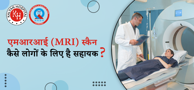
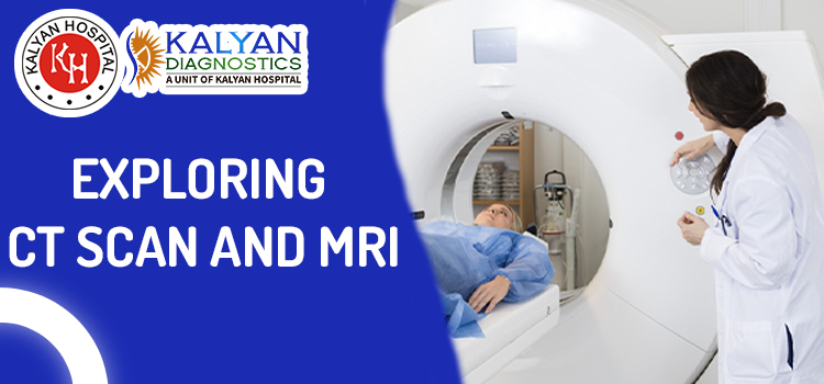
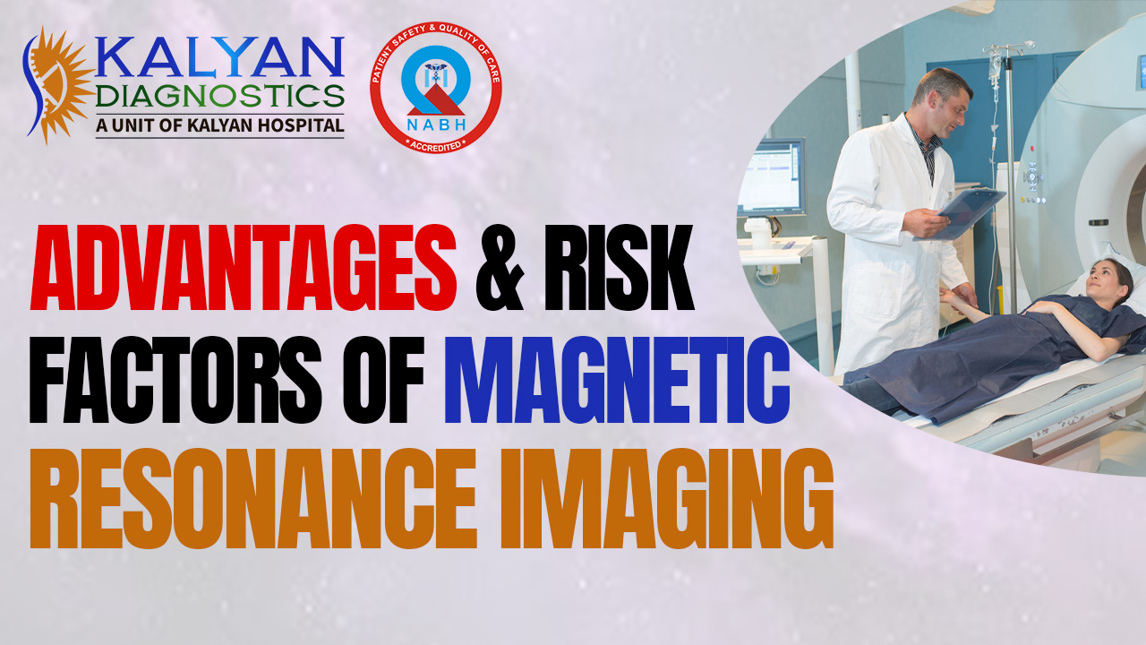
The MRI machine is a long, narrow tube that is openable from both ends. For taking the MRI X-ray, a person has to lie down on a table that slides into the opening of the tube; a technician monitors the MRI machine and you from the other room by using a microphone. 

Things you have to tell your doctors before the MRI Scan

Many people have a fear of enclosed spaces. A condition of phobia of enclosed spaces is known as claustrophobia. Your doctors suggest you take steroids in order to feel relaxed and less anxious. 

The procedure of MRI scans. 

The MRI machines create the strongest magnetic force around you. The radio waves are directly emitted into your body. The MRI procedure is painless. People do not feel the magnetic and radio waves. There are no moving parts around the individuals. MRI scans can affect the internal part of the magnet, producing repair tapping, thumping, and other kinds of noises. Your doctors suggest wearing earplugs and listening to music just to block the noise. A contrast material, gadolinium, will be injected into the intravenous line into a vein in the hand and arm. The material of contrast is lucrative for making certain details clearer. Gadolinium causes allergic reactions in some cases. It helps pinpoint the portions of your brain that control these actions. For the best and safest test, visit the best Diagnostic center in Ludhiana.

Benefits of MRI Scan

MRI scans can be beneficial for taking images of the different parts of the body. It takes images of the Head, joints, abdominal area, and other parts from different directions. Your tissues become less contrasty in MRI than in the CT scan and other different imaging scans. These images provide information to physicians and can be useful in diagnosing a wide variety of diseases and conditions.

Risk factor of the MRI scans. 

The images are made without the use of ionizing radiation. Patients do not see the harmful effects of ionizing radiation. But while there are no known health hazards from temporary exposure to the MR environment, the MR environment involves a strong, static magnetic field, a magnetic field that changes with time (pulsed gradient field), and radiofrequency energy, each of which carries specific safety concerns:

  • The strong, static magnetic field will attract magnetic objects (from small items such as keys and cell phones to large, heavy items such as oxygen tanks and floor buffers) and may cause damage to the scanner or injury to the patient or medical professionals if those objects become projectiles. Careful screening of people and objects entering the MR environment is critical to ensure nothing enters the magnet area that may become a projectile.
  • The magnetic fields that change with time create loud knocking noises, which may harm hearing if adequate ear protection is not used. They may also cause peripheral muscle or nerve stimulation that may feel like a twitching sensation.
  • The radiofrequency energy used during the MRI scan could lead to heating of the body. The potential for heating is greater during long MRI examinations.

The diagnostic center does the MRI with a prescription that is given by a doctor who is well-qualified and experienced. Book an appointment with Kalyan Diagnostics for better-quality scans, and it is also one of the best centers for MRI Test in Ludhiana.
A Comprehensive Guide To Get Best Mri Services In Ludhiana

When a patient is suffering from any condition that affects the internal organs of humans or they might have damage done to the soft tissues in their body, medical professionals advise them to get either a CT scan or MRI done.


If you are wanting to get any of these procedures done to you and want to know the MRI cost in Ludhiana, then you should read this blog post.


KALYAN DIAGNOSTICS Ludhiana is famous for procedures like CT scan and MRI. Their team of expert doctors like Dr.Rajinder Singh has cured many patients till today. 


It was established to provide the best medical care at economical prices. They are equipped with the most advanced equipment and use them to get precise diagnoses every time.


At KALYAN DIAGNOSTICS, you can get the best CT Scan Cost in Ludhiana. A large number of patients are treated at the hospital each day successfully. 


MRI uses elements like magnetic fields and also computer-generated radio waves to generate expansive imagery of internal organs and tissues in the human body. 


Images obtained during MRI are vital in understanding the condition of soft tissues, which in turn helps to understand and diagnose conditions like brain tumor, cancerous tumours and other kinds of issues like brain disorders.


KALYAN DIAGNOSTICS provides the best MRI services in Ludhiana. They have a well-trained team of medical professionals who are experts in their fields.


They are an all-encompassing institute that provides all diagnostic facilities under one roof like Ultrasound, Digital X-Ray, Doppler, Digital OPG etc.


They are widely famous for providing accurate, reliable, affordable diagnostic services at minimal costs.


They are open all throughout the day, all days of the week, so you can visit the hospital at any time you require. 


We also provide emergency admission at any time. You can directly come to our hospital without the requirement of a prior booking, this gives the patients a better chance to get admitted in case of a health emergency.


They are also present online. One can visit their official website to get detailed information about the institution and the services they provide. This makes it easier for the patients to be well-versed in the hospital.


At KALYAN DIAGNOSTICS, MRI is performed using state-of-art technology and well-experienced doctors. An MRI machine is a tube-like magnetic machine; when a patient lies down inside the MRI machine, the magnetic field of the machine temporarily aligns itself to the water molecule present in the body of the patient.

Once aligned, the atoms release feeble signals which are used to create cross-sectional imagery of the patient’s body.


It is a non-invasive procedure, meaning it does not require any intentional cuts and cruises to the patient.


Both MRI and CT scans are done differently. While a Ct scan uses X-Ray, MRI depends upon a strong magnetic field to obtain imagery of the internal organs. Mri is usually not the first choice for the diagnosis; MRI is done when a CT scan is not able to detect an issue.


THINGS TO CONSIDER BEFORE GETTING MRI DONE 

a)Presence of metal- If a patient has any type of metal implants in his/her body, they should let the doctor know about it. A patient with a heart pacemaker should never get an MRI done, it can lead to harmful consequences. 


b)Pregnancy- Although the effect of MRI on an unborn child is still not known, you should tell the doctor if you are pregnant or if you think you might be pregnant.


c)Care for children-  Usually, children are hesitant to go inside the MRI machine, they are given anti-anxiety medications, so if you are concerned, you should tell the doctor.


d)feeling of claustrophobia- Some patients are claustrophobic, they are uncomfortable inside the MRI machine, they should tell the doctor, the doctor might give you medication for anxiety.

How to prepare for MRI Scans?

For the different problems, there are different scans and tests. The different types of tests tell you about the different problems that can occur in your body. MRI is a type of scan and stands for Magnetic resonance imaging. MRI is a type of diagnostic test that can create detailed images of nearly every structure and organ inside the body. MRI uses magnets and radio waves to produce images on a computer. MRI does not use ionizing radiation. Images produced by an MRI scan can show organs, bones, muscles and blood vessels. 

What is the definition of MRI? 

Most of the people will only ever hear the MRI referred to by its abbreviation. The Full form of MRI is Magnetic Resonance imaging. In this test, Radio Waves are used, and powerful magnets are used to click the image from the depth of the human body. These images send the correct and critical information about each and everything that is going on inside your body. The best diagnostic center in Ludhiana is Kalyan Diagnostic.                                                                                                                                                                                                                         

How do you have to prepare for the MRI scans? 

Before going for MRI Scans you have to tell everything about your medical history and medical conditions to your doctor. 

If you have claustrophobia, tell your doctor before going for an MRI scan. 

For the MRI scan, you have to lie down in a tube-like machine. The length of time you will spend in the machine will vary. It could be as long as an hour. If you have claustrophobia, you are going for an MRI Scan, which will surely cause discomfort and sometimes anxiety to the patients. For better cooperation, tell your doctor about your problem so that they can manage some for the treatment of your phobia. The center for the scan of MRI in Ludhiana is well known. 

Do not take your jewelry with you at home. 

Wearing metal is one of the top items on the list of what not to do before an MRI. Because an MRI is essentially a giant magnet, the MRI techs will ask you to remove any metal items from your person before entering the machine, including any jewelry you may be wearing. If you’re wearing inexpensive pieces, this may not be a cause for concern.

Tell all your corners to your doctor, honestly.

Tell your doctor if in your body any piece of metal is implanted because the MRI Scans need high magnetic power. If there is any metallic implantation, then tell your doctor before the scans. The metallic implants include kidney history, pacemaker, injury that can be adjusted with metal, and many more. Tell your health care professionalist before the MRI scans. It causes problems during the scanning procedure. 

Benefits of MRI Scan

MRI scans can be beneficial for taking images of the different parts of the body. It takes images of the Head, joints, abdominal area, and other parts from different directions. Your tissues become less contrasty in MRI than in the CT scan and other different imaging scans. These images provide information to physicians and can be useful in diagnosing a wide variety of diseases and conditions.

Risk factor of the MRI scans. 

The images that are made without the use of ionizing radiations. Patients do not meet the harmful effects of ionizing radiation. But while there are no known health hazards from temporary exposure to the MR environment, the MR environment involves a strong, static magnetic field, a magnetic field that changes with time (pulsed gradient field), and radiofrequency energy, each of which carries specific safety concerns:

  • The strong, static magnetic field will attract magnetic objects (from small items such as keys and cell phones to large, heavy items such as oxygen tanks and floor buffers) and may cause damage to the scanner or injury to the patient or medical professionals if those objects become projectiles. Careful screening of people and objects entering the MR environment is critical to ensure nothing enters the magnet area that may become a projectile.
  • The magnetic fields that change with time create loud knocking noises, which may harm hearing if adequate ear protection is not used. They may also cause peripheral muscle or nerve stimulation that may feel like a twitching sensation.
  • The radiofrequency energy used during the MRI scan could lead to heating of the body. The potential for heating is greater during long MRI examinations.

The diagnostic center does the MRI with a prescription that is given by a doctor who is well-qualified and experienced. Book an appointment with Kalyan Diagnostics for better-quality scans.

एमआरआई (MRI) सर्वाइकल स्पाइन करवाने के क्या है लागत, परिणाम व सम्पूर्ण जानकारी ?

एमआरआई (MRI) जोकि हर बीमारी को पकड़ने में काफी मददगार साबित होती है। साथ ही एमआरआई की मदद से हम अंधरुनि समस्या को जानकर उसका इलाज अच्छे से करवा सकते है। वहीं एमआरआई से सर्वाइकल स्पाइन का इलाज व जाँच को कैसे किया जाता है, इसके बारे में आज के लेख में चर्चा करेंगे ;

एमआरआई (MRI) कैसे सहायक है सर्वाइकल स्पाइन की जाँच के लिए ?

  • एमआरआई सर्वाइकल स्पाइन रीढ़ के सर्वाइकल क्षेत्र (गर्दन के पीछे की तरफ) की जांच और पता लगाने में मदद करता है। यह गर्दन और रीढ़ की अंदरूनी चोट या फ्रैक्चर का पता करने में मदद करते है। 
  • यदि आपको गर्दन के क्षेत्र में दर्द है तो डॉक्टर आमतौर पर इसकी सलाह देते है। यह परीक्षण गर्दन और सर्वाइकल स्पाइन के कोमल ऊतकों का 3-डी दृश्य देता है। वही लुधियाना में MRI का खर्च या सर्वाइकल स्पाइन की कीमत 3000 रुपये से लेकर 6000 रुपये तक है।

सर्वाइकल स्पाइन के MRI में क्या शामिल हो सकते है ?

  • सात कशेरुक जो C1 से C7 तक होते है। 
  • रक्त वाहिकाएं। 
  • नसे। 
  • पट्टा। 
  • बंधन। 
  • उपास्थि आदि इसके MRI में शामिल होते है। 

क्या है सर्वाइकल MRI स्कैन ? 

  • सर्वाइकल एमआरआई (मैग्नेटिक रेजोनेंस इमेजिंग) गर्दन के पीछे की हड्डियों, जिन्हें सर्वाइकल स्पाइन कहा जाता है, की विस्तृत छवियां उत्पन्न करने के लिए एक चुंबकीय क्षेत्र और रेडियो तरंगों का उपयोग करते है।  
  • परीक्षण के दौरान, एमआरआई मशीन व्यक्ति के शरीर में एक अस्थायी चुंबकीय क्षेत्र बनाती है, जो शरीर में मौजूद हाइड्रोजन परमाणुओं को क्षेत्र के साथ संरेखित करने के लिए मजबूर करती है।
  • अगर आप गर्दन के पीछे की हड्डियों या सर्वाइकल स्पाइन की समस्या से परेशान है, तो इससे बचाव के लिए आपको लुधियाना में बेस्ट रेडियोलॉजिस्ट से अपनी जाँच को करवाना चाहिए।

सर्वाइकल स्पाइन MRI का उद्देश्य क्या है ?

  • रीढ़ की हड्डी में चोट लगी हो तो इसके लिए इस स्कैन का चयन किया जाता है।  
  • हड्डियों या कोमल ऊतकों में ट्यूमर या कैंसर होने पर भी आप इस स्कैन का चयन कर सकते है। 
  • हर्नियेटेड डिस्क होने पर इस स्कैन का चयन करना।
  • डिस्क फलाव की समस्या होने पर इसका चयन करना। 
  • जन्मजात या शारीरिक जन्म दोष होने पर इस स्कैन का चयन करना।
  • हड्डियों की बनावट में असामान्यता के आने पर इसका चयन करना।
  • स्पाइनल स्टेनोसिस की समस्या होने पर इसका चयन करना। 
  • ऊपरी अंगों में अकड़न होने पर इसका चयन करना।

MRI सर्वाइकल स्पाइन के विभिन प्रकार क्या है ?

  • WSS के साथ MRI का सर्वाइकल स्पाइन करवाना। 
  • एमआरआई सर्वाइकल स्पाइन फ्लेक्सन-एक्सटेंशन। 
  • ब्रेन जांच के साथ एमआरआई सर्वाइकल स्पाइन की जाँच। 
  • मल्टीपल स्केलेरोसिस का पता लगाने के लिए। 
  • ALS के पहचान के लिए। 
  • गतिशील एमआरआई सर्वाइकल स्पाइन।
  • कंट्रास्ट के साथ एमआरआई सर्वाइकल स्पाइन। 

सर्वाइकल स्पाइन MRI क्यों किया जाता है ?

  • गर्दन, बांह या कंधे में गंभीर दर्द जो इलाज के बाद भी कम ना हो तो इस जाँच का चयन करें। 
  • पैर में कमजोरी या सुन्नता जैसे लक्षणों के साथ गर्दन में दर्द होने पर। 
  • रीढ़ की हड्डी के जन्म दोष या रीढ़ की हड्डी में चोट का पता लगाना। 
  • रीढ़ की हड्डी में संक्रमण या ट्यूमर का पता लगाने के लिए। 
  • रीढ़ की हड्डी में गठिया का पता लगाने के लिए। 
  • परीक्षण स्पाइन सर्जरी से पहले या बाद में भी किया जा सकता है।

सर्वाइकल MRI कैसे किया जाता है ?

  • रेडियोग्राफर आपको स्कैन कक्ष में प्रवेश करने से पहले डेन्चर और विग सहित सभी धातु की वस्तुओं को हटाने का निर्देश देंगे। 
  • वे आपको हॉस्पिटल गाउन पहनने के लिए कह सकते है। 
  • आपको एक बिस्तर पर लेटना होगा, जो एमआरआई स्कैनर मशीन के अंदर चला जाएगा। 
  • प्रक्रिया में एक डाई (कंट्रास्ट) की आवश्यकता हो सकती है, जो परीक्षण से पहले आपकी बांह में एक नस के माध्यम से दी जाएगी। 
  • कंट्रास्ट कुछ क्षेत्रों को अधिक साफ-साफ देखने में मदद करता है। 
  • आपको बिल्कुल स्थिर होकर लेटना होगा, क्योंकि हरकतें तस्वीरों को धुंधला बना सकती है। 
  • परीक्षण आमतौर पर 30-60 मिनट तक चलता है लेकिन इसमें अधिक समय भी लग सकता है। 

सर्वाइकल MRI के जोखिम क्या है ?

  • कंट्रास्ट से एलर्जी की प्रतिक्रिया को करना, जोकि काफी जोखिम भरा होता है। 
  • एमआरआई मशीन में मौजूद मजबूत चुंबक पेसमेकर और अन्य धातु प्रत्यारोपण में खराबी या शरीर के अंदर हिलने-डुलने का कारण बन सकते है। 
  • उपयोग किया जाने वाला कंट्रास्ट उन किडनी रोगों वाले लोगों के लिए हानिकारक हो सकता है जिन्हें डायलिसिस की आवश्यकता होती है।

सुझाव :

सर्वाइकल स्पाइन में अगर आपको दिक्कत है तो इस समस्या का सामना करने के लिए आपको एमआरआई का चयन करना चाहिए, क्युकी एमआरआई की मदद से ये जानना काफी आसान हो जाता है की व्यक्ति को किस तरह की समस्या है। 

इसके अलावा आप चाहें तो इस टेस्ट को कल्याण डायग्नोस्टिक्स हॉस्पिटल से भी करवा सकते है। 

एमआरआई (MRI) स्कैन स्पाइन लम्बर यानि (पीठ में होने वाले दर्द) के मामलों में कैसे है सहायक ?

एमआरआई (MRI) स्कैन जोकि हर तरह की समस्या का स्पष्ट रूप में मूल्यांकन करते है साथ ही इसका चयन करके हमें सब कुछ स्पष्ट हो जाता है की हम किस तरह की बीमारी से जूझ रहें है और साथ ही एमआरआई (MRI) स्कैन डॉक्टरों के लिए काफी सहायक माना जाता है। इसके अलावा एमआरआई स्पाइन लम्बर की समस्या को कैसे विस्तार से बाहर लाने में हमारी मदद करेंगे इसके बारे में हम सम्पूर्ण लेख में चर्चा करेंगे ;

क्या है एमआरआई (MRI) स्कैन ?

  • MRI (मैग्नेटिक रेसोनेंस इमेजिंग) एक जांच की प्रक्रिया है। इसमें शरीर के अंदर की उच्च गुणवत्ता वाली, विस्तृत तस्वीरें बनाने के लिए एक बड़े चुंबक, रेडियो तरंगों और कंप्यूटर का उपयोग किया जाता है। 
  • एमआरआई जांच का चयन अगर कोई भी करता है, तो उसे दर्द की समस्या का सामना नहीं करना पड़ता, इसलिए इस स्कैन का चयन बच्चे भी कर सकते है। 

इस स्कैन का चयन करने से बेशक आपको दर्द की समस्या का सामना नहीं करना पड़ता, लेकिन जरूरी है की अगर आप इस स्कैन का चयन करने जा रहें है तो इससे पहले लुधियाना में MRI का खर्च कितना है इसके बारे में जरूर जानकारी हासिल करें।

एमआरआई (MRI) स्कैन से क्या-क्या बाते सामने आती है ?

  • एमआरआई स्कैन का चयन जब आप करते है तो आपको मस्तिष्क, रीढ़, जोड़ों, हृदय, लीवर, और कई अन्य अंगों जैसे शरीर के कोमल-ऊतक व अन्य संरचनाओं का पता एमआरआई (MRI) चित्रों के द्वारा आसानी से पता लग जाता है। 
  • कैंसर, हृदय की बीमारी, मांसपेशियों और हड्डियों की बीमारियों, और कई अन्य बीमारियों का डायग्नोसिस एमआरआई स्कैन से किया जा सकता है। इस स्कैन की मदद से हम रीढ़ की हड्डी व पीठ की हड्डी में दर्द की समस्या का भी पता लगा सकते है।

एमआरआई (MRI) स्पाइन लम्बर स्कैन में किस तरह की तकनीक का प्रयोग किया जाता है 

  • चुंबकीय अनुनाद इमेजिंग (एमआरआई) स्कैन एक सामान्य प्रक्रिया है, जिसका उपयोग रीढ़ की हड्डी और अन्य अंगों के निदान के लिए भी किया जाता है। 
  • इस प्रक्रिया में चुंबकीय तरंगों का इस्तेमाल रीढ़ की हड्डी के विस्तृत चित्र का निर्माण करने के लिए उपयोग किया जाता है। 
  • इसमें मरीज को मेज़ पर लेटा कर एमआरआई मशीन के अंदर ले जाया जाता है, जो, एक बड़े डोनट की तरह लगता है। 
  • एमआरआई स्कैन आक्रामक नहीं है और यह एक दर्द रहित प्रक्रिया है। 
  • वहीं एक्स-रे और सीटी स्कैन के विपरीत, एमआरआई स्कैन में विकिरण का उपयोग नहीं होता है, इसलिए यह एक सुरक्षित प्रक्रिया है।

एमआरआई (MRI) स्पाइन लम्बर के प्रकार क्या है ?

  • एमआरआई स्पाइन लम्बर के लिए कंट्रास्ट एजेंट शरीर में इंजेक्शन या मौखिक रूप से दिया जाता है। कंट्रास्ट एजेंट आमतौर पर बेहतर छवियाँ देता है, जबकि बिना कंट्रास्ट इसके विपरीत है। हालांकि, आपके डॉक्टर यह फैसला करते है कि आपको कौन-से टेस्ट के लिए जाना है।
  • अधिकांश एमआरआई स्कैन स्पाइन लम्बर में रोगी को लिटाकर मशीन में ले जाने की आवश्यकता होती है। जो लोग बंद मशीन में जाने से डरते है, उनके लिए यह एक चिंता का विषय हो सकता है। तकनीशियन उन मामलों में एनिस्थिसिया दे सकते है या वह ओपन एमआरआई का विकल्प भी सामने वाले मरीज को दे सकते है, पर ध्यान रहें ये स्कैन थोड़ा मेहगा है।
  • टेस्ला चुंबकीय शक्ति की माप की एक इकाई है। एमआरआई मशीन 1.5 टेस्ला या 3 टेस्ला हो सकती है। 3 टेस्ला एमआरआई स्कैन मशीन तेज गति से बेहतर गुणवत्ता की छवियों का उत्पादन कर सकती है। हालांकि, यह अधिक महंगी होती है। इसके अतिरिक्त, भारत में हो रहे अधिकांश एमआरआई स्कैन के लिए 1.5 टेस्ला मशीन पर्याप्त है।
  • स्पाइन लम्बर क्षेत्र के लिए आवश्यक जानकारी के आधार पर, डॉक्टर स्क्रीनिंग या सामान्य एमआरआई की सिफारिश कर सकते है। 

ध्यान रहें अगर आपको स्पाइन लम्बर करवाना है, तो इसके लिए आप लुधियाना में बेस्ट रेडियोलॉजिस्ट का चयन करें।

स्पाइन लम्बर स्कैन को क्यों कराया जाता है ?

  • पीठ के निचले हिस्से में लगातार दर्द अगर हो तो आप इस स्कैन का चयन कर सकते है। 
  • रीढ़ की हड्डी के निचले भाग में चोट लगने पर। 
  • मस्तिष्क या रीढ़ की हड्डी में कैंसर के लक्षण का पता लगाने पर। 
  • रीढ़ की हड्डी में जन्म से कोई समस्या होने पर। 
  • पैरों के सुन्न पड़ने पर। 
  • पीठ के निचले हिस्से में सर्जरी होने पर।

सुझाव :

अगर आप पीठ या रीढ़ की हड्डी में दर्द की समस्या से परेशान है तो इससे बचाव के लिए आपको स्पाइन लम्बर स्कैन को जरूर करवाना चाहिए, वहीं आप चाहें तो इस स्कैन को कल्याण डायग्नोस्टिक्स हॉस्पिटल से भी करवा सकते है।

भारत में क्या है एमआरआई (MRI) स्कैन की लागत व पूरी प्रक्रिया ?

कई बार व्यक्ति को कुछ ऐसी बीमारियां हो जाती है कि उसे पता ही नहीं चलता है की वो किस तरह की बीमारी का सामना कर रहा है पर ऐसे में एमआरआई (MRI) स्कैन एक बेहतरीन विकल्प माना जाता है अंदरूनी बीमारी को जानने में तो अगर आप भी किसी तरह की बीमारी का सामना कर रहे और वो बीमारी आपको काफी परेशान कर रही है तो उससे निजात पाने के लिए आपको एमआरआई स्कैन का चयन करना चाहिए ;

क्या है एमआरआई (MRI) स्कैन ?

  • चुंबकीय अनुनाद इमेजिंग यानि “मैग्नेटिक रेज़ोनेंस इमेजिंग” एक चिकित्सा इमेजिंग तकनीक है जो कि आपके शरीर में अंगों और ऊतकों की विस्तृत छवियां बनाने के लिए चुंबकीय क्षेत्र और कंप्यूटर से उत्पन्न रेडियो तरंगों का उपयोग करती है। वहीं यह एक्स-रे (विकिरण) का उपयोग नहीं करता है।
  • दूसरी और अधिकांश एमआरआई मशीनें बड़ी, ट्यूब के आकार की चुम्बक होती हैं। वही जब आप MRI मशीन के अंदर लेटते हैं, तो चुंबकीय क्षेत्र अस्थायी रूप से आपके शरीर में पानी के अणुओं को पुन: बनाता है।

 एमआरआई (MRI) स्कैन करवाने से क्या पता चलता है ?

  • मस्तिष्क, रीढ़, जोड़ों, हृदय, लीवर, और कई अन्य अंग जैसे शरीर के कोमल-ऊतक संरचनाओं की एमआरआई (MRI) चित्रों के द्वारा ज्यादा सटीक रूप से पहचाना जा सकता है। कैंसर, हृदय की बीमारी, मांसपेशियों और हड्डियों की बीमारियों, और कई अन्य बीमारियों का डायग्नोसिस एमआरआई स्कैन से किया जाता है।

 एमआरआई (MRI) स्कैन का उपयोग कहां किया जाता है ?

  • मस्तिष्क और रीढ़ की हड्डी की असामान्यताएं को जानने के लिए।
  • शरीर के विभिन्न भागों में द्रव्यमान, ट्यूमर, अल्सर और अन्य विसंगतियों की उपस्थिति को जानना।
  • पेट में हृदय, यकृत और अन्य अंगों से संबंधित स्थितियों को जानना।
  • फाइब्रॉएड और एंडोमेट्रियोसिस जैसी महिलाओं में पेल्विक दर्द से संबंधित स्थितियों को जानना।
  • बांझपन के मामले में महिलाओं में संदिग्ध मूत्र असामान्यताओं का मूल्यांकन करना।

आप एमआरआई से भी इन परेशानियों की जाँच करवा सकती है या आप लुधियाना में CT स्कैन का खर्च कितना आएगा, इसके बारे में जान के भी इसकी जाँच सीटी स्कैन से करवा सकते है।

एमआरआई (MRI) स्कैन को करवाने में खर्चा कितना आता है ?

  • सिटी स्कैन, एमआरआई की जांच के लिए मरीज से 500 से लेकर 8 हजार या उससे ज्यादा की फीस ली जाती है। हालांकि ये टेस्ट अभी कॉन्ट्रैक्ट बेस पर मशीनें लगाकर करवाई जा रही है।
  • इसके अलावा मरीज़ के अंगों पर भी इसका खर्चा निर्भर करता है की वो शरीर के किस अंग की जाँच करवाने जा रहा है। वही लुधियाना में MRI का खर्च जगह और हॉस्पिटल कहाँ स्थित है इस पर भी पड़ता है।

एमआरआई (MRI) स्कैन करवाने के लिए बेस्ट हॉस्पिटल व सेंटर ?

  • अगर आपको अपने शरीर के अंधरुनि अंगों की जाँच अच्छे से करवानी है तो इसके लिए आप हमारे  डायग्नोस्टिक्स सेंटर का चयन कर सकती है।

क्या है एमआरआई (MRI) स्कैन की प्रक्रिया ?

  • रोगी को स्कैन के दौरान अस्पताल का गाउन पहनने की आवश्यकता होती है। साथ ही, स्कैन के दौरान आभूषण या धातु पहनने की अनुमति नहीं है।
  • एमआरआई स्कैन एक गैर-इनवेसिव और दर्द रहित प्रक्रिया है। और अवधि स्कैन किए जा रहे शरीर के हिस्से के आकार और कितनी छवियों को लेने की आवश्यकता के आधार पर 15 मिनट से एक घंटे या उससे अधिक तक समय लग सकता है।

निष्कर्ष :

उम्मीद करते है की आपको पता चल गया होगा की आपको कब और किस समय एमआरआई (MRI) स्कैन करवाना चाहिए।

जानिए चेहरे का मेकअप कैसे MRI रिपोर्ट में लेकर आया कैंसर की समस्या को !

MRI जिसको हमारे शरीर के जाँच के लिए एक बहुत ही बढ़िया मशीन माना जाता है, इसकी मदद से आपके शरीर का अच्छे से जाँच किया जाता है। पर क्या आप जानते है की अगर आप इस जाँच को मेकअप लगा कर करवाने जाते है तो इसकी रिपोर्ट गलत भी आ जाती है, वहीं कुछ मामलों में तो मेकअप के बाद MRI को करवाने से कैंसर जैसे मामले भी सुनने को मिले है, तो चलिए जानते है की आपको MRI के दौरान किन बातों का ध्यान रखना चाहिए, ताकि जाँच के बाद आपकी रिपोर्ट गलत न आए ;

क्या है MRI जाँच ?

  • चुंबकीय अनुनाद इमेजिंग यानि (मैग्नेटिक रेज़ोनेंस इमेजिंग) एक चिकित्सा इमेजिंग तकनीक है जो कि आपके शरीर में अंगों और ऊतकों की विस्तृत छवियां बनाने के लिए चुंबकीय क्षेत्र और कंप्यूटर से उत्पन्न रेडियो तरंगों का उपयोग करती है। वहीं यह मशीन एक्स-रे का उपयोग नहीं करता है।
  • अधिकांश मामलों में हमने देखा है की एमआरआई मशीनें बड़ी, ट्यूब के आकार की चुम्बक होती है। जब आप MRI मशीन के अंदर लेटते है, तो चुंबकीय क्षेत्र अस्थायी रूप से आपके शरीर में पानी के अणुओं को पुन: संरेखित करने लगते है। 
  • इसके अलावा रेडियो तरंगें इन संरेखित परमाणुओं को बेहोश सिग्नल उत्पन्न करने का कारण बनती है, जिनका उपयोग क्रॉस-अनुभागीय एमआरआई छवियों को बनाने के लिए किया जाता है।

जानिए क्या है ओपन और कंट्रास्ट वाला एमआरआई (MRI) ?

  • ओपन MRI की बात करें तो ये एक खुला एमआरआई मशीन के प्रकार को संदर्भित करता है जो छवियों को लेता है। आम तौर पर, एक खुली एमआरआई मशीन में आपके ऊपर और नीचे दो फ्लैट मैग्नेट होते है, जिनके बीच में आपको लेटने के लिए एक बड़ी जगह होती है। यह दो तरफ खुली जगह की अनुमति देता है और क्लॉस्ट्रोफोबिया (claustrophobia) को कम करता है। 
  • फिर बात करें कंट्रास्ट MRI की जो कुछ एमआरआई परीक्षाएं कंट्रास्ट सामग्री के इंजेक्शन का उपयोग करती है। कंट्रास्ट एजेंट में गैडोलीनियम होता है, जो एक दुर्लभ पृथ्वी धातु है। और जब यह पदार्थ आपके शरीर में मौजूद होता है, तो यह आस-पास के पानी के अणुओं के चुंबकीय गुणों को बदल देता है, जिससे छवियों की गुणवत्ता बढ़ जाती है।

इन दोनों में से किसी भी MRI का चयन करने से पहले लुधियाना में MRI का खर्च कितना है, इसके बारे में जरूर जानकारी हासिल करें।

एमआरआई (MRI) जाँच से क्या पता चलता है ?

  • आपके मस्तिष्क और आसपास के तंत्रिका ऊतक का पता चलना।
  • आपकी छाती और पेट के अंग, जिसमें आपका हृदय, लीवर, पित्त पथ, गुर्दे, प्लीहा, आंत्र, अग्न्याशय और अधिवृक्क ग्रंथियां नज़र आती है।
  • स्तन के ऊतक का भी पता चलता है।
  • आपके रीढ़ की हड्डी में लगें चोट का भी इसके द्वारा पता चलता है।
  • आपके मूत्राशय और प्रजनन अंगों सहित श्रोणि अंग में विकार के बारे में भी इस टेस्ट के माध्यम से जानकारी आपको हासिल हो सकती है।
  • रक्त वाहिकाओं में परेशानी हो तो उसके बारे में भी इस टेस्ट के माध्यम से पता लगाया जा सकता है।
  • लसीकापर्व (प्रतिरक्षा प्रणाली के भाग) की भी जाँच की जा सकती है MRI की मदद से।

MRI जाँच से सही बीमारी का पता तभी लगाया जा सकता है, जब आप इस जाँच को लुधियाना में बेस्ट रेडिओलॉजिस्ट से करवाते है।

क्या MRI जाँच का चयन करने से व्यक्ति कैंसर की बीमारी से ग्रस्त हो जाता है ?

  • आपको बता दें कि एमआरआई एक दर्दरहित प्रोसिजर होता है, जिसमें 15 से 90 मिनट तक का समय आराम से लग जाता है। वहीं इस टेक्नॉलजी में शक्तिशाली चुंबकों, रेडियो वेव और एक कम्प्यूटर का इस्तेमाल होता है, जिससे शरीर के अंदर की पूरी जांच की जाती है। इसकी मदद से डॉक्टर घाव, रक्त की नसों में डैमेज, कैंसर या फिर दिल की बीमारियों का भी पता लगा सकते है।
  • जहां एक ओर डॉक्टर और लैब असिस्टेंट एमआरआई स्कैन से पहले मरीज के शरीर से मेटल हटाने के लिए कहते है, वहीं कभी-कभी वह चेहरे के मेकअप को नजरअंदाज कर देते है, जिसमें धातु तत्व होते है। जिस वजह से ही जांच रिपोर्ट गलत आ जाती है।
  • इसलिए जरूरी है की अगर आप MRI की जाँच करवाने जा रहें है तो ध्यान रहें की आपके शरीर में मेटल की कोई भी चीज न हो और न ही आपके चेहरे पर मेकअप लगा होना चाहिए। 
  • तो MRI से जाँच की रिपोर्ट कभी गलत नहीं आती, बस कुछ हमारे द्वारा और कुछ डॉक्टरों के द्वारा बातें ध्यान न होने की वजह से इसकी रिपोर्ट गलत आ जाती है। और घबराए न MRI की जाँच को करवा कर आप कैंसर पीड़ित नहीं हो सकते।

सुझाव :

एमआरआई (MRI) एक बहुत ही सुरक्षित और दर्द रहित जाँच है, जिसको करवाने से आप कैंसर पीड़ित नहीं होते, बल्कि ये आपके शरीर की समस्या के बारे में जाँच करते है और आपकी बीमारी को खुल कर सबके सामने लाते है।

वहीं अगर आप MRI की जाँच को सुरक्षित तरीके से करवाना चाहते है, तो इसके लिए आप कल्याण डायग्नोस्टिक्स हॉस्पिटल का चयन कर सकते है। 

निष्कर्ष :

MRI जाँच एक बहुत ही बेहतरीन जाँच प्रक्रिया है, जिसको करवा कर आप अपने शरीर में उत्पन्न हुई बीमारी का जड़ से खात्मा करने में काफी सहायक साबित होते है। इसके अलावा अफवाओं पर यकीन न करें की इस जाँच का चयन करने से व्यक्ति की समस्या और बढ़ सकती है, बल्कि इससे जुडी किसी भी तरह की समस्या का आपको सामना करना पड़ रहा है, तो इससे बचाव के लिए आपको अनुभवी रेडिओलॉजिस्ट डॉक्टर के पास आना चाहिए।

जानिए एमआरआई (MRI) से हम कैसे स्तन कैंसर का पता लगा सकते है ?

एमआरआई (MRI) स्कैन एक ऐसे स्कैन की पद्यति में गिना जाता है, जिसकी मदद से हर बीमारी का पता आसानी से लगाया जा सकता है। इसलिए तक़रीबन डॉक्टरों के द्वारा भी तरह की अंधरुनि समस्या होने पर इस स्कैन का चयन करने के लिए कहा जाता है। वहीं आज के लेख में जानेगे की हम कैसे स्कैन के माध्यम से स्तन कैंसर का पता लगा सकते है ;

महिलाओं में क्या है स्तन या ब्रेस्ट का कैंसर ?

  • महिलाओं में कैंसर से होने वाली मौत में सबसे ज्यादा ब्रेस्ट कैंसर जिम्मेदार माना जाता है। ऐसे में अगर समय रहते या समय से पहले इस बीमारी की पहचान हो जाए तो होने वाली मौतों की संख्या को कम किया जा सकता है। 
  • वहीं काफी अनुसंधानकर्ताओं के द्वारा ये पता चला है की मरीजों के ब्रेस्ट में हेल्दी सेल्स यानी कोशिकाओं की तुलना कैंसर ग्रस्त कोशिकाओं और गैर कैंसरकारी कोशिकाओं से की जाती है।
  • स्तन कैंसर, महिलाओं के स्तनों के अंदर विकसित होने वाला एक तरह का कैंसर है। जो आमतौर पर महिलाओं में आम है, लेकिन आजकल यह पुरुषों में भी हो सकता है। यह ज्यादातर मामलों में एक या एक से अधिक गांठों के रूप में भी शुरू होता है, जिसे स्तनों के अंदर महसूस किया जाता है। वहीं यह गांठें स्तन के ऊपरी या नीचे के हिस्सों में पाया जाता है।

एमआरआई (MRI) की मदद से महिलाओं के स्तन कैंसर का पता कैसे लगा सकते है ?

  • कैंसर एक घातक बीमारी है और महिलाओं में ब्रेस्ट कैंसर या अन्य समस्याएं होना भी आजकल बहुत सामान्य होते जा रहा है। ब्रेस्ट कैंसर और ऐसे अन्य गंभीर रोगों का पता लगाने के लिए ब्रेस्ट का एमआरआई करवाया जाता है। इस स्कैन टेस्ट में कैंसर की पूरी स्थित यानी कौन-से स्टेज का कैंसर है ये पता चल जाता है। 
  • हम ये भी कह सकते है कि इस टेस्ट का प्रयोग उन महिलाओं में जिनमे कैंसर होने की संभावना बहुत अधिक होती है उनकी स्क्रीनिंग टूल के रूप में किया जाता है। ब्रेस्ट एमआरआई स्कैन में डॉक्टर को रोगी की ब्रेस्ट टिश्यू की साफ तस्वीर मिलती है, जिससे वो महिला में ब्रेस्ट कैंसर के बारे में अधिक सही से जान पाते है। 
  • ब्रेस्ट एमआरआई डॉक्टर को कैंसर के आकार और कैंसर कितना फैल चुका है, इन सब चीजों को माप कर कैंसर के चरण को निर्धारित करने में भी मदद मिलती है। 
  • ब्रेस्ट एमआरआई में किसी भी तरह की आयनीकृत रेडिएशंस का प्रयोग नहीं होता। डॉक्टर इनका प्रयोग महिलाओं में स्तन के ऊतकों का प्रशिक्षण करने के लिए करते है। जिनमें कैंसर का खतरा अधिक है उनमें इस बीमारी के शुरूआती लक्षण दिखाई देने लगते है। 
  • कैंसर चाहे किसी को भी हो सब में कुछ न कुछ लक्षण दिखाई जरूर देते है। इसलिए जरूरी है उन लक्षणों के नजर आते ही डॉक्टर से मिले। और स्तन कैंसर को अंदेखा न करें।
  • स्तन कैंसर का रिजल्ट बेहतर आए इसके लिए जरूरी है की सबसे पहले आप इस बीमारी की जाँच को लुधियाना में बेस्ट रेडियोलॉजिस्ट से जरूर करवाए।

स्तन कैंसर का चयन किन स्थितियों में किया जाता है ? 

  • कैंसर के निदान के बाद स्तनों में अतिरिक्त ट्यूमर या हानिकारक टिश्यू को खोजने के लिए।
  • 25 वर्ष से कम उम्र की महिलाओं में स्तन ऊतक का पता लगाने के लिए।
  • हैवी टिश्यू की जांच करने के लिए।
  • मैमोग्राम या अल्ट्रासाउंड जैसे अन्य इमेजिंग परीक्षणों के परिणामों की पुष्टि करने के लिए।
  • गर्भवती या स्तनपान कराने वाली महिलाओं में स्तन के ऊतकों का आकलन करना।
  • कीमोथिरेपी की प्रभावशीलता की जांच के लिए।
  • उन महिलाओं की जांच के लिए जिनकी पुनर्निर्माण सर्जरी हुई हो।
  • अधिकांश महिलाएं नियमित स्कैन के रूप में ब्रेस्ट एमआरआई से नहीं गुजरती है। जिन लोगों को ब्रेस्ट कैंसर होने की संभावना अधिक होती है। उन लोगों के लिए डॉक्टर ब्रेस्ट एमआरआई के साथ एक मैमोग्राम को शुरुआती जांच उपकरण के रूप में जोड़ सकते है। 
  • स्तन कैंसर का एमआरआई करवाने से पहले लुधियाना में MRI का खर्च कितना है, इसके बारे में जरूर जानकारी हासिल करें।

महिलाओं में स्तन कैंसर के क्या लक्षण है ?

  • स्तन या बाँह के नीचे गांठ या मोटापन जैसी समस्या का सामना करना। 
  • स्तन के आकार या आकृति में बदलाव का आना। 
  • स्तन की त्वचा में ढीलेपन या सिकुडेपन जैसी समस्या का सामना करना। 
  • स्तन नलिका से अधिक स्तन दूध के अलावा कुछ और निकलना। 
  • कंधे के नीचे गांठ जैसी सूजन का महसूस होना।
  • अगर आपको भी ऐसा महसूस हो या आपमें भी उपरोक्त लक्षण नज़र आए तो इससे बचाव के लिए आपको कल्याण डायग्नोस्टिक हॉस्पिटल का चयन करना चाहिए, लेकिन ध्यान रहें लक्षणों को कृपया नज़रअंदाज़ न करें।

सारांश :

इसके अलावा स्तन कैंसर में एमआरआई (MRI) क्या भूमिका निभाते है या ये कैसे महिलाओं के कैंसर को बाहर निकालने में मददगार साबित होते है, इसके बारे में हम इस छोटे से लेख में चर्चा कर चुके है। वहीं एमआरआई स्तन कैंसर ही नहीं बल्कि हर अंधरुनि बीमारी का पता करने में सहायक साबित होते है।